Scoliosis: Is “High Side of the Rainbow Adjusting” Really Enough?
 Saturday, February 12, 2011 at 5:16PM

Dr. Joe Betz, B.S., D.C
ICA Board Member,
PCCRP Board Member
Private Practice Boise, ID
Idaho Chiropractic Association Board Member
CBP Instructor
INTRODUCTION
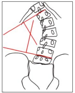
Scoliosis is the most common “spinal deformity” that presents to a chiropractic office. Although “scoliosis” is traditionally defined as a “lateral curvature of the spine” (as seen in Figure 1), it is more appropriately described in terms of its 3-dimensional configuration, including coronal and sagittal plane changes.
As a student I became interested in learning about scoliosis and how chiropractors manage this common condition. However, when I attended Chiropractic College, practically all of my clinical training concerning management of scoliosis patient could be summed up in the phrase: “Adjust on the ‘high side of the rainbow”. I assumed this was woefully inadequate and with the guidance of Dr. Don Harrison, I began investigating the background of the biomechanics of scoliosis and began to learn the application of Chiropractic Biophysics methods in the management of the scoliotic spine.
Don Harrison taught that in order to effectively manage scoliosis, you must first have a thorough understanding of the underlying biomechanics of the structural scoliosis. I read prominent research articles and textbooks, dating back to the mid 1800’s, and worked forward to present studies. To say that there is a significant amount of literature on the topic is a dramatic understatement. I still have boxes full of the thousands of articles I collected on the topic.
One thing that attracted me to the “old” articles was their extensive knowledge of the 3-dimensional nature of the scoliosis deformity. Also, the scoliosis practitioners of the late 1800’s and early 1900’s didn’t have “braces” (in modern day terms) nor did they have surgical techniques. They used exercises and what could be described as traction methods. They only abandoned these methods in the 1940’s with the development of the lucrative Milwaukee braces and what has proven to become ever-evolving, and even more lucrative surgical procedures.


The Sagittal Plane
The sagittal configuration of the spine has been of interest to scoliosis researches for many years. It is commonly accepted that when you view the true lateral image of the apex of a thoracic scoliosis on an X-ray, described in 1965 as the plan d’ election view1, a lordosis will always be present…yes a “thoracic lordosis”. Many spine doctors have described this and corresponding non-surgical (and non-bracing) methods many years ago.2 With a thorough understanding of the 3-D nature of scoliosis, the sagittal alignment of the scoliotic spine becomes increasingly important.
Whereas the lordotic nature of the thoracic spine (at the scoliosis apex) is well understood, the sagittal presentation of the lumbar spine has less congruency. This is likely due to the varying measurement methods used to determine extent of lumbar lordosis, including flawed assessment procedures such as a flexible ruler or spinal pantograph.
The implication of scoliosis on the sagittal alignment of the cervical spine is well established. In 1975 Winter, etal3 reported a loss of lordosis in the cervical spine in scoliotic patients with associated loss of thoracic kyphosis. However, they reported no data. The authors did state, “thoracic lordosis was usually accompanied by a compensatory cervical kyphosis”.
In 1989, Cruikshank, et al4 reasoned that an area of lordosis, for example in the thoracic spine, must be balanced by a kyphosis above and below. This is what Hilibrand et al5 found in their excellent study on the sagittal configuration of the cervical spine in idiopathic scoliosis patients. They report, “we found the greatest cervical kyphosis in patients with thoracic hypokyphosis”. Most recently in 2009, Morningstar and Stitzel6 found similar results.
So, although the effect of the 3-D scoliotic deformity is most pronounced in the thoracic and lumbar regions, there are consequences for the sagittal cervical spine as well. However, these changes in cervical curvature are most certainly secondary to the thoracic scoliosis (and lordosis), NOT a primary etiological factor.
SUMMARY
An understanding of what a scoliosis actually looks like in a real patient is the most crucial aspect of clinical decision making when managing these patients. The biggest mistake clinicians make is relying upon 2-D representations of the deformity on radiographs without understanding the actual 3-D configuration of the spine itself.
For decades, CBP® doctors have the goal of restoring normal spinal alignment in three dimensions. Scoliosis is a mechanical problem. CBP® provides a rational biomechanical explanation for effective clinical treatment of the scoliotic deformity. Any chiropractors interested in managing scoliosis more successfully should consider learning CBP® technique.
Dr. Deed Harrison and I will be teaching two scoliosis seminars on CBP® management of scoliosis in 2011:
- April 9-10 in NY/NJ, and
- August 13-14 in Seattle.
For details and to register visit www.idealspine.com or call CBP Seminars at 800-346-5146.
REFERENCES
- Peloux J du, Fauchet R, Faucon B, Stagnara P. Le plan d’election pour l’examen radiologique des cyphoscolioses. Rev Chir Orthop 1965; 51:517-524.
- Steindler A. Diseases and deformities of the spine and thorax. St Louis, CV Mosby Company, 1929.
- Winter RB, Lovell WW, Moe JH. Excessive thoracic lordosis and loss of pulmonary function in patients with idiopathic scoliosis. J Bone Joint Surg [Am] 1975;57:972-6.
- Cruickshank JL, Koike M, Dickson RA. Idiopathic scoliosis in three dimensions. J Bone Joint Surg [Br] 1989:71:259-63.
- Hilibrand AS, Tannenbaum DA, Graziano GP, Loder RT, Hensinger RN. The sagittal alignment of the cervical spine in adolescent idiopathic scoliosis. J Spinal Dis 1995;15:627-32.
- Morningstar MW, Stitzel CJ. The Relationship between cervical kyphosis and idiopathic scoliosis. J Vertebral Subluxation Res. October 13, 2008; 1-4.
 CBP Seminars | Comments Off |

