Entries from April 1, 2010 - April 30, 2010

Tuesday
Apr132010

HOW TO FIND GREAT STAFF

clip_image002

Eric Huntington, DC

Co-Owner Developer of the Chiropractic Business Academy

While there are many aspects to a successful practice, one key element is hiring the right staff. At the Chiropractic Business Academy we provide chiropractors with the skills and procedure necessary to not only find quality staff, but to also train those staff to become super effective team members. In fact, one of the reasons that we are so popular with our clients is because WE TRAIN THEIR STAFF!

It all Starts with Finding the Right Person to Hire

You have to confront the fact that a great majority of the people who will come to your office to interview are not employable. From my experience of owning a large staff run office for ten years and the hundreds of chiropractors we consult every week, I estimate that on average, one in thirty people that you interview are employable—and about one in one hundred are that super star that will help bring your practice to the next level. You may get lucky and find a great person in the first five or ten, but realize that you were fortunate, and don’t stress if you really have to search for your next one.

So, this means that you have to set up your hiring strategy so that it can manage large numbers of people without disrupting the rest of your practice. Here is one way to do that.

Interview every week, even if you don’t necessarily need to hire someone right now. You always want to be on the lookout for that one in one hundred. If you find that super star, you can always find a place for them which will grow your practice. I recommend to my clients that a marketer, if well trained and productive, will always make you more money than they cost you. So marketing is one place you can put an “extra” person.

When we are fully staffed, we only use free advertising mediums on the internet or flyers on cork boards around town. When we really need new staff, we pay for newspaper ads, etc. Always keep a file of decent prospects if you can’t hire them right away.

Hold a group interview at the same day and time each week. Have a dedicated phone number that goes to a voicemail which nobody picks up (you can use a cell phone for this as well). On the recording, leave the day and time of the interview—so if it’s always the same, you never have to change it. On the message be sure to leave all necessary data--address, directions, etc. Remind them not to leave a message.

Train your receptionist to have employment applications on clipboards with pens ready on the day of your interview-- as people often come early. You do the first part of the interview in a group. Tell the whole group your name and that you will be meeting with them briefly. Starting with the first person who is done with their application (or the first person who walked in, if you don’t care whether or not they finish the application) peel them off from the group, somewhere that is semi-private.

If your office space allows, don’t take them into a room, as you’ll find yourself getting trapped and wasting time. In my office, we can have them walk out of the reception seating area and meet the interviewer next to the front desk-- They are literally standing for this part of the interview.

Three things that I look for in this short interview are,

  1. Do they communicate well: Can you understand what they are saying—volume, accent, properly structured sentences, etc? When they answer questions, are their answers truly answers to your questions, or is it sort of off the topic? When they originate something, is it appropriate to the setting and is it consistent with the conversation?
  2. Do they present well: Did they dress appropriately? Are they well groomed—as opposed to dirty or messy? Did you feel comfortable with them near you and would you feel comfortable that they could help you handle something really important to you?
  3. Are they positive: Are they cheerful, excited about life and an opportunity to work?

There are other very important things to look for which take more extensive training to learn. These things are covered in our client workshops and courses.

For those people that you do not wish to have back for a second interview, tell them that you are reviewing applications and will only be contacting those that seem to be a good fit for the position, and have them back for a second interview—tell the person that you’ll make those phone calls within a few days. Thank them for coming to the office, shake their hand and end it quickly—but very politely. A key to getting yourself to do this weekly is not wasting time.

For those that you think might be a good candidate, have a short test on hand which you can have them do right in the office-- on that first interview.

If you’d like the test I use, you can call into The Chiropractic Business Academy and ask for Brian. He can give you the test I use.

Call 888-989-0855

The Next Step is a Working Interview for Your Best Candidates! To learn how to conduct this interview visit our website and read the rest of this article. www.ChirobizAcademy.com

Tuesday
Apr132010

Audits, Accounting and Problems

clip_image002

R.J. Hammett, DC

Chances are that at some time in your practice life, you’re going to be hassled. An insurance company won’t like you, a fellow chiropractor will turn you into the board, you’ll do or say something stupid. Whatever the case, some “authority” will come knocking at your door. Trust me, it is not a good experience. So what do you do? Well, prevent defense is the best game plan. It won’t solve all your problems or totally protect you, but it may give you a fighting chance. By the way, if you’re doing something illegal or unethical, nothing will save, so quit now and stand up right.

Here is a check list of things you should look over:

1. Are you HIPPA compliant?

2. Do you use informed consent on all patients?

3. Do you “really” listen to your patients and write down what is said and done?

4. Do you re-x-ray or re-exam every patient every 15th-30th visit or so?

5. Do you re-examine those who just ‘drop in’ for a quick adjustment?

6. Do you review your outgoing billing at least twice a year? Do your codes match your documentation?

7. Are you keeping up on continuing education courses?

8. Do you know when to refer a non-responding patient?

9. Are you answering patients concerns?

10. Are your staff members working with you or against you?

11. Do you examine/x-ray the parts you adjust or treat?

12. Do you watch your promotions i.e : “Guarantee a cure” practicing outside your licence law?

13. Are you using outregous techinques or other on the edge stuff in your clinic?

14. Are you keeping up with changes in the laws of your state?

Ok, you have done everything right but someone wants a piece of you? What do you do?

1. Never, ever talk to any investigator from the board or person without the presense of an attorney. Ignore the inclination to cooperate fully until you have legal representation.

2. Do not grant an entry. Do not turn over patient records or other office material, send them to your attorney to send to any requesting authority.

3. Do not allow your staff to be interviewed.

4. Do not allow any authoriziation into your office without an attorney present.

5. Be respectful and polite, and tell the investigator that you will cooperate through your attorney.

6. The investigator is NOT your friend.

7. If you receive a subpoena, verify it with your attorney first.

8. Talk to a lawyer that is specialized in your type of case; NOT a general practice attorney.

I hope this helps, in fact, I hope you never need this information. But remember, bureaucracies are not your friend, nor will they ever be. Their job is to police, let them do their jobs, you do yours.

Til next time. RJH

Monday
Apr122010

Restoration of an Abnormal Cervical Lordosis Using the DENNEROLL: A CBP® Case Report

clip_image002

clip_image004

clip_image006

Brian Paris, BS, DC Deed E. Harrison, DC

Advanced CBP Fellow

Private Practice Rockville, MD

President CBP Seminars, Inc.

Vice President CBP Non-Profit, Inc.

Chair PCCRP Guidelines

Editor—AJCC

Introduction

In surgical1,2 and non-surgical literature3-5, it has been found that patients with less cervical lordosis have statistically significant increases in neck, upper thoracic, and shoulder pain and likely overall poorer health outcomes. The following case report suggests that the loss of cervical lordosis, forward head carriage, spinal arthritis and disc disease (S.A.D.D.), with concomitant sympomatology is consistent with current literature relating to cervical lordosis and pain.1-5

Case History Key Features

A 59 year old female present to the practice of one of the authors (BP) seeking help for recurrent and chronic dizziness, neck and shoulder pain and stiffness, left arm tingling, sleep deprivation, and generalized fatigue. Her initial visit was on 10-7-2009 and she indicated that the current episode of her ailments had been present for at least the previous 2 months.

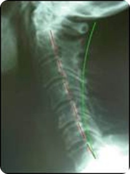
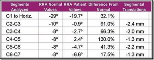
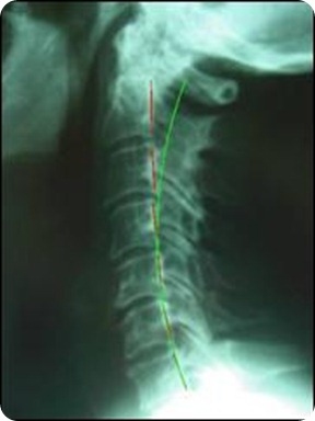
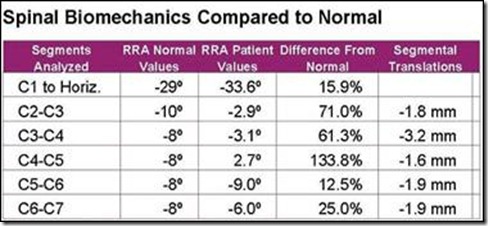
Cervical radiographs consisting of a neutral lateral and anterior-posterior were obtained. The lateral cervical radiograph revealed signs of spinal arthritis and disc disease (S.A.D.D) increasing in severity at the C5-C7 levels with possible rheumatoid changes in the upper cervical region. The patient had a large forward head carriage (58 mm from C2-C7) and a 70% reduction of the normal cervical lordosis. Radiographic analytical measurements and comparison to normal values were performed with the PostureRay® computerized software system from PostureCo. See Figure 1.

clip_image008clip_image010

Figure 1. Patient Pre-Lateral Cervical Radiograph and segmental alignments relative to ideal values. The green semi-circular curvature is the ideal curvature proposed by Harrison et al and the dashed red line represents the path of the patient’s posterior vertebral bodies and visually depicts the amount of displacement. The initial cervical lordosis demonstrated a 70% reduction, 12.6° from C2-C7 using the Harrison Posterior Tangent method.6,7 The initial forward translation was 58 mm; using posterior superior body corner of C2 relative to a vertical line originating at the posterior inferior body corner of C7.

CBP Mirror Image® Interventions

The patient was recommended and agreed to a treatment plan of spinal correction using CBP Technique mirror image adjustment, exercise, and traction methods. The treatment frequency was 3 times per week for 40 visits over approximately 13 weeks. The Patient presented to and actively participated at all appointments. Each visit consisted of mirror image adjusting, mirror image exercises and the Denneroll cervical orthotic to improve the cervical lordosis and reduce abnormal posture displacements.

  • Mirror Image Adjustments

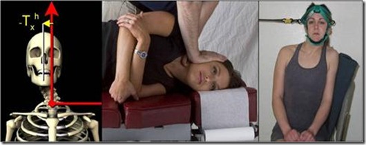
Beginning on 10-9-09, the patient was administered mirror image adjustments to correct Right Head Translation and Anterior Head Translated postures. See Figures 2 and 3.

  • Mirror Image Exercises

Active rehabilitative care was administered to the patient beginning on 10-9-09. Since the patient’s abnormal posture was found to be Right Head Translation (-TxH) and Anterior Head translation (+TzH), the patient began mirror image exercises in left head translation to correct right head translation and posterior head translation without extension due to dizziness. See Figures 2 and 3.

clip_image013

Figure 2. The patient presented with the abnormal posture of Right Head Translation (-TxH). Mirror image adjustments were given in Left Head Translation. Mirror image exercises in Left Head Translation were given to the patient as part of CBP Active Rehabilitative care.

clip_image016

Figure 3. The patient presented with the abnormal posture of Anterior Head Translation (+TzH). Mirror image adjustments were given in Posterior Head Translation with extension to improve the lordosis and reduce forward head translation. Mirror image exercises in Posterior Head Translation (extension exercises caused increased dizziness) were given to the patient as part of CBP Active Rehabilitative care.

  • The Denneroll Cervical Orthotic Intervention

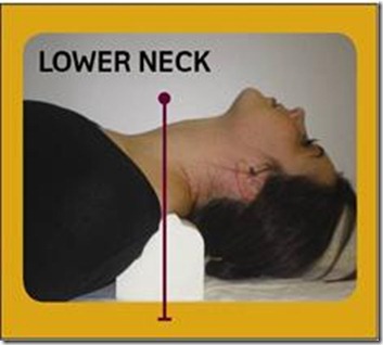
The patient experienced considerable difficulty in performing passive and active cervical extension. Accordingly, the more advanced types of CBP in office traction methods could not be performed by the patient.1 Thus, the Denneroll cervical orthotic device (Figure 4) was provided to the patient as the sole method of in office cervical corrective traction-stretching. Denneroll (adult small size) corrective stretching began on 10/19/2009 and continued for a total of 34 treatment sessions. Patient time started at 3 minutes per session and then increased up to 10 minutes per Denneroll session each visit in the office.

For the current patient, the Denneroll was placed in the upper thoracic/lower cervical region (Figure 4). This placement of the Denneroll will cause significant posterior head translation, will increase the upper thoracic curve if the large device is used (this is why the small device was used herein), and will increase the overall cervical lordosis.

clip_image019

Figure 4. Denneroll corrective orthotic application in the lower cervical region. This lower neck placement is for abnormal cervical curvatures having:

· Normal or a mild loss of the upper thoracic kyphosis;

· Loss of the mid-lower cervical curve;

Anterior head translation of approximately ≤ 40mm

clip_image022clip_image024

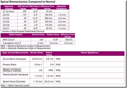
Figure 5. Patient Post-Lateral Cervical Radiograph and segmental alignments relative to ideal values. The green semi-circular curvature is the ideal curvature proposed by Harrison et al and the dashed red line represents the path of the patient’s posterior vertebral bodies and visually depicts the amount of displacement. The Follow-up cervical lordosis demonstrated a 56% reduction, 18° from C2-C7 using the Harrison Posterior Tangent method.6,7 The initial forward translation was 19 mm; using posterior superior body corner of C2 relative to a vertical line originating at the posterior inferior body corner of C7.

Re-Examinations:

In addition to the initial examination, 2 follow-up evaluations were performed over the course of the 40 sessions. At each examination, structural and functional responses to care were evaluated and patient symptoms were recorded and monitored using the Neck Disability Index and the Rand 36- Health Status Questionnaire. See Table 1. The brief examination findings are summarized here:

  • 11/12/2009: Decreased dizziness; thinking more clearly; energy level (fatigue) same; left arm tingling improvingànow only intermittent;
  • 12/14/2009: Infrequent bouts of dizziness, only occasional tingling in left arm;
  • 1/13/2010: No reports of dizziness, occasional tingling in left arm.
  • Overall Improvements: Significant health improvements were noted by patient since beginning treatment: “No dizziness”; “more clear-headed”. At most recent follow-up, dizziness had not returned. Only occasional tingling in left hand was reported and the patient has elected to continue with a 2nd phase of CBP Corrective Care.

Table 1. Initial and Follow-up Neck Disability and Rand-36 Questionnaire results.

Questionnaire

Initial Exam 11-12-2009

Re-Exam 1-13-2010

Neck Disability

22% Pain interference with ADL’s

12% Pain interference with ADL’s

Rand-36-Health Status Questionnaire

12-14-09 1-13-10

Physical Function

60

75

Social Function

62.5

75

Role Physical

100

100

Role Emotional

0

100

Mental Health

64

72

Energy-Fatigue

45

55

Pain

45

67.5

Health Perception

77

52

Discussion

The Denneroll orthotic applies a passive 3-point bending force to the cervical spine that is generally well tolerated and is most consistent with the Pope-2-way type of in office corrective traction force.6The Denneroll is available in 2 adult sizes (adult large and adult small) and the adult small device was used for the present patient. The Denneroll size and placement of the device must be consistent with both the shape of the cervical curve and the amount/type of sagittal head translation correction that is desired for the given patient.

In the current case report, the combination of CBP mirror image methods resulted in improvement of cervical spine vertebral subluxation towards normal alignment. The Denneroll Orthotic for cervical lordosis corrective traction-stretching was the only type of CBP Traction utilized herein. Thus, it appears that the Denneroll Orthotic device assisted in cervical spine correction and improvement in chronic patient symptoms, disability, and altered health when applied in combination with mirror image adjustments and active mirror image exercises.

We will continue to test the Denneroll device in appropriate patient cases and provide the results in future articles.

References

  1. Lowery G. Three-dimensional screw divergence and sagittal balance: a personal philosophy relative to cervical biomechanics. Spine: State of the Art Reviews 1996;10:343-356.
  2. Kawakami M, Tamaki T, Yoshida M, Hayashi N, Ando M, Yamada H. Axial symptoms and cervical alignments after cervical anterior spinal fusion for patients with cervical myelopathy. J Spinal Disorders 1999;12:50-56.
  3. Kai Y, Oyama M, Kurose S, et al. Traumatic thoracic outlet syndrome. Orthop Traumatol 1998;47:1169-1171.
  4. Harrison DD, Harrison DE, Janik TJ, Cailliet R, Haas JW, Ferrantelli J, Holland B. Modeling of the Sagittal Cervical Spine as a Method to Discriminate Hypo-Lordosis: Results of Elliptical and Circular Modeling in 72 Asymptomatic Subjects, 52 Acute Neck Pain Subjects, and 70 Chronic Neck Pain Subjects. Spine 2004; 29:2485-2492.
  5. McAviney J, Schulz D, Richard Bock R, Harrison DE, Holland B. Determining a clinical normal value for cervical lordosis. J Manipulative Physiol Ther 2005;28:187-193.
  6. Harrison DE, Harrison DD, Haas JW. CBP Structural Rehabilitation of the Cervical Spine. Chapters 2 & 6. CBP Seminars, 2002; pgs:147-151. ISBN 0-9721314-0-X.
  7. Harrison DD, et al. Spine 2004; 29:2485-2492.

image


Monday
Apr122010

PostureRay: Mirror Image® Traction Links Here We Come!

clip_image002

clip_image004

Joseph Ferrantelli, DC

Private Practice New Port Richey, FL

CTO, CBP® Seminars

CEO, PostureCo

Many Chiropractors have successfully begun using PostureRay®in their practices and have provided us feedback on how it has aided in growing the practice by reducing the time for objective and reliable x-ray analysis, as well as generating patient friendly Report of Findings marketing materials, not to mention the multifunctional digital view box.

Well now we have some more exciting news! We have just begun the process of programming the most requested of additional modules, that being, linking a patient’s x-ray findings with mirror image® traction protocols and set up procedures. This module will be aimed at reducing mistakes in traction selection when considering the all pertinent x-ray biomechanical findings. For example, occasionally we receive calls from Chiropractors to our CBP hotline where they have pursued traction treatment for a patient with a cervical kyphosis, adjusted them, and exercised them, all with only minimal improvement in their post x-ray findings. Now to a less experienced structural based doctor, one might jump to the conclusion that CBP methods have failed. However, more often than not, it was merely an incorrect mirror image traction setup for the patient, or worse yet, the wrong traction method may have been administered.

Consider the sagittal cervical spine as an example where many patient cases have altered thoracic alignment coupled with the abnormal cervical lordosis. Mechanically, the subluxated thoracic kyphosis would change the choice and set up for the patient’s cervical spine traction. Unfortunately, many chiropractors do not routinely obtain lateral thoracic x-rays which would limit the chances for the patient’s success. There are at least 16 categories of abnormal cervical curvatures encompassing the type upper thoracic kyphosis and the type of sagittal plane head posture. In relationship to these categories there are 4 -primary types of in office CBP Traction equipment with each having several types of possible setups specific to the curve and posture. Furthermore there are now 3 types of viable home traction units. Thus, choosing the appropriate mirror image traction application type and setup for a specific lateral cervical subluxation is not a 'simple task'.

clip_image006

clip_image008

clip_image010

clip_image012

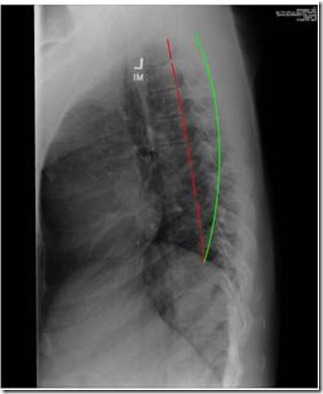
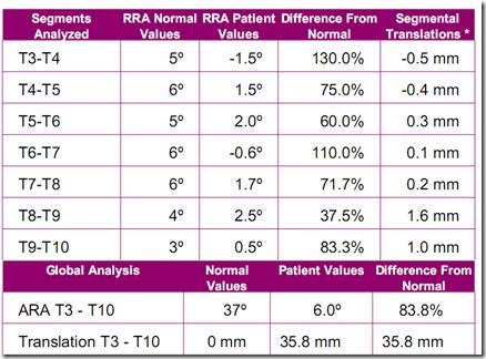
Consider the case in Figure 1 (lateral cervical) and Figure 2 (lateral thoracic). Looking at the cervical curvature we can identify:

· A straightened lower cervical lordosis with a ‘flattened’ or near vertical C7-T1 posterior vertebral body alignment,

· A cervical kyphosis from C2-C5 segments—mid cervical kyphosis if you will,

· A numerical value of C2-C7 anterior translation is found and seen, however, this is not true head translation. The reason C2 is anterior to C7 is due to the cervical kyphosis from C2-C5,

· There is a mild flexion of the skull on the atlas.

Now, looking at the thoracic curvature we can identify:

· Anterior translation of T3 relative to T10,

· A nearly straight thoracic kyphosis showing an 83.8% reduction in curvature.

Clinical Questions Concerning This Case:

1. What is the appropriate type of cervical curve traction to be used in the office?

2. What would the exact setup look like for this type of in office traction?

3. What is the appropriate type of home orthotic traction to be administered in this case?

4. What would the exact setup look like for this type of home orthotic?

Solutions

In the near future, PostureRay will be able to guide you to the likely most appropriate traction types and setups. In cases like these, the treating Chiropractor can become frustrated, especially in higher volume settings, and worse yet - the patient may remain subluxated due to ineffective interventions. The result may be that the patient and doctor are left believing chiropractic may not be able to ‘correct’ their condition and that only symptomatic care is appropriate.

With this new module under development, under direction of Dr. Deed Harrison, we aim to remove as much guess work as possible, suggesting better set up protocols with both angles of pull, patient positioning, and of course, choice of appropriate traction methods based on patient specific x-ray analysis, conditions and symptomatic ailments.

Look for this additional module to become available towards the end of this year!

If you have questions about how PostureRay can help your practice, feel free to visit our website atwww.postureco.com or email us at sales@postureco.com or direct questions to myself atdrjoe@idealspine.com. Always remember, a digital x-ray machine is NOT required to utilize PostureRay®.


Monday
Apr122010

Leg Length Inequality: Recent Studies & A New Full Length Shoe Lift Orthotic

clip_image002

Deed E. Harrison, DC

President CBP Seminars, Inc.

Vice President CBP Non-Profit, Inc.

Chair PCCRP Guidelines

Editor—AJCC

Introduction

The assessment of the patient with observed postural distortion and spine disorders would seem incomplete without the analysis of a weight bearing radiographic view of the pelvis and lumbar spine, including the femur heads. One of the more common structural findings in patients with lumbar spine disorders is the presence of an anatomical leg length inequality (ALLI). See Figure 1 for Postural Consequences of the ALLI. Although there is a significant body of literature discussing the epidemiology of ALLI, considerable controversy still exists over ALLI’s significance in patient populations with spinal disorders.1,2

In the CBP Lumbar Rehabilitation Textbook (2007),3 we presented a contemporary and complete review of the literature pertaining to ALLI and its biomechanical consequences, appropriate analysis, and interventions. In this current report, I would like to present a few recent investigations delineating some important findings regarding ALLI and its conservative treatment with lift intervention.

Postureclip_image006

Figure 1. Typical Abnormal Posture Caused by a Left Anatomical Short Leg. In A (left photo), for a left ALLI, the thoracic cage will translate to the left side and laterally flex to the right. Often, the pelvis will translate slightly towards the side of ALLI (left in this case). In B (right photo), the pelvis will rotate anterior on the short leg side. If the shoulders and feet are aligned parallel, this means that the thorax has rotated posterior in the exact degrees that the pelvis rotated anterior. In other words, the thorax typically rotates posterior on the short leg side.

Recent Study #14

In a 2010 prospective study of 3026 patients 50-79 yrs of age, Harvey et al.4 sought to determine the correlation of ALLI to prevalent, incident and progression of knee osteo-arthritis (OA). They4 measured ALLI using full leg x-rays. Their findings are striking:

• ALLI ≥ 1 cm was associated with prevalent and symptomatic OA of knee. OR = 1.9

• The shorter leg had increased incident symptomatic and progressive OA of the Knee. OR = 1.5 & 1.3.

Conclusion:Radiographic LLI was associated with prevalent, incident symptomatic, and progressive Knee OA. LLI is a potentially modifiable risk for Knee OA.”4

Recent Study #25

In a 2009 cohort study of 3012 people, Golightly and colleagues5 examined the relationship between chronic Knee and/or hip symptoms to ALLI. In 2006 of the subjects, there was an ALLI of ≥ 2 cm. Subjects in their sample with ALLI were statistically more likely to have knee symptoms (p<0.001). After adjusting for several confounding variables, knee symptoms were statistically associated with the presence of an ALLI (OR= 1.41 p< 0.001).

They5 stated, “LLI was moderately associated with chronic knee symptoms and less strongly associated with hip symptoms. LLI may be a new modifiable risk factor for therapy of people with knee or hip symptoms” .5

Orthotic Correction of ALLI Improves Chronic Pain

In a 2005 randomized trial, Defrin et al6 found statistically significant improvements in chronic low back pain in patients receiving shoe lifts compared to no treatment in the control subjects. In both the control and lift treatment groups, the measured LLI was between 5-10mm as measured via pelvic radiographs. This important study provides evidence that patients with chronic low back pain with concomitant ALLI between 5-10 mm may benefit from orthotic intervention aimed at leveling the sacral base and femur head heights. They stated,

Shoe inserts appear to reduce CLBP and functional disability in patients with LLDs of 10mm or less”.

In 2007, Golightly et al7 reported on improvements in twelve (6 males and 6 females) consecutive chronic low back pain patients between the ages of 19-62 years of age with radiographic ALLI between 6.4-22.2 mm. Subjects were treated with orthotic intervention determined by the height that best relieved patient symptoms. Using the visual analog scale and disability questionnaires, statistically significant reductions in chronic pain intensity and disability scores were found.

clip_image009

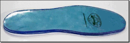
Figure 2. Sole Supports full‐foot lifts are made of an easily cleanable, non‐compressible vinyl. If you are a Sole Supports certified provider, you receive a discounted rate for the full foot lift in combination with a Custom Orthotic order. However for non-Sole Supports certified providers, CBP Seminars has arranged to be a distributor for these innovative, contemporary, and biomechanically sound full-foot lifts. See www.idealspine.biz for details and ordering information.

New Full Length Shoe Lift for ALLI Correction From Sole Supports

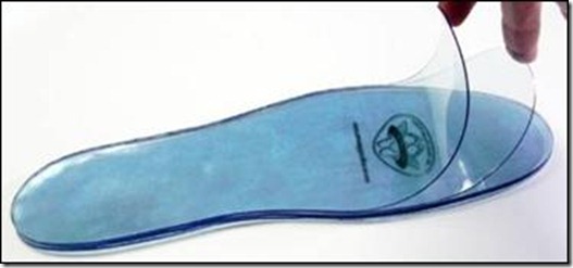
An exciting product has just been launched on the market by Sole Supports as a new option for treatment of anatomical leg length inequality (ALLI). See Figure 2. This product is a full‐foot lift, made of non‐compressible vinyl and is available in 1mm peel able increments. Each lift consists of six, one‐millimeter layers of vinyl that allow you to adjust the height of the lift to suit your patient’s needs. See Figure 3.

clip_image012

Figure 3. Sole Supports Full Foot Lift. Each lift consists of six, one‐millimeter layers of vinyl that allow you to adjust the height of the lift to suit your patient’s individual needs. They are available in a range of both men’s and women’s sizes. The 6 mm size was chosen as a limit of what will generally fit in the average shoe.

Biomechanical Benefits of Full-Length Foot Lift

Problematically, the standard heel lift has a gradual taper being larger at the heel and smaller towards made foot. Though these tapered heal lifts have been found to be effective at relieving patients chronic pain syndromes, biomechanically there can be consequences to the functionality of the foot-ankle complex over long-term use.

clip_image015

Figure 4. Different types of shoe lifts available. Note that all of these lifts have a strong increased heel height and taper towards the mid-foot. Theoretically this taper may alter forefoot forces and heel-achilles tendon tightness due to ankle plantar flexion.

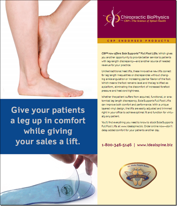
In contrast, use of the new full foot lift allows for correction of ALLI without changing ankle angulation or increasing plantar flexion of the foot; thus not compromising proper foot biomechanics. In theory, with a full‐foot lift, the foot will remain level (not considering the slope of the shoe itself) and the leg is lifted as a platform. The full foot lift would also limit changes in forefoot pressure and heel-achilles tendon tightness that can result because of ankle plantar flexion due to a tapered lift. These Sole Support full‐foot lifts should be used to address both acquired or functional leg length discrepancies, as well as anatomical discrepancies.

Editor’s Note: Sole Supports has graciously licensed CBP Seminars to distribute their innovative, contemporary, and biomechanically sound full-foot lifts. See www.idealspine.biz for details and ordering information.

References

  1. Knutson GA. Chiropractic & Osteopathy 2005;13(11):1-10.
  2. Brady RJ, Dean JB, Skinner MT, Gross MT. JOSPT 2003; 33:221-234.
  3. Harrison DE, Harrson DD, Haas JW, Oakley PA. Chapter 4. CBP Structural Rehabilitation of the Lumbar Spine. Evanston, WY: Harrison CBP Seminars, Inc., 2007.
  4. Harvey et al. Assoc of leg-length inequality with knee osteoarthritis. An Intern Med 2010; 152:287-95

5. Golightly et al. Symptoms of knee and hip in individuals with and without LLI. Osteoarthritis Cartilage 2009; 17: 596-600.

  1. Defrin R, et al. APMR 2005;86:2075-2080
  2. Golightly YM, Tate JJ, Burns CB, Gross MT. J Orthop Sports Phys Ther. 2007; 37(7):380-388.

image


Monday
Apr122010

Can You Prove “Soft Tissue Injuries”?

clip_image002

clip_image004

Matthew D. Powell,

Board Certified Civil Trial Attorney - Florida

Matthew Powell is a board certified civil trial lawyer in Tampa, Florida, who started his career representing over 20 insurance companies. After learning the defense side, he quickly found he could not represent insurance companies and started out on his own to represent injured victims. He has had the honor of obtaining $1,000,000 verdicts for his clients. He can be reached at matt@EPTInjuryLaw.com.

Soft tissue injuries are the most common types of injuries that chiropractic physicians treat. As a personal injury trial lawyer, I have had many opportunities to represent clients who suffered these permanent and debilitating injuries. Almost every trial lawyer will tell you, that “soft tissue” types of cases are the hardest and most difficult cases to win at trial. You may ask, why are soft tissue injuries so hard to prove?

Let’s consider what type of tissue “soft tissue injuries are”. First, the most common tissues are muscles, ligaments, and tendons. But what about nerves, the spinal cord, the brain, our heart, our organs, and our skin? When you think about it, most of the human body is comprised of soft tissue.

How do we, as trial lawyers prove soft tissue injuries? The answer is, by objectifying, or visually documenting, and demonstrating soft tissue injuries in such a way that the jurors can see the injury. Most jurors are very skeptical, however, their skepticism is won over easily when they can see objective proof of an injury. Today’s jurors are familiar with such shows as CSI, and expect high tech, sophisticated, scientific evidence that will prove the claim. And the good news is, that this type of evidence is readily available, easy to use, and produces a great impression upon jurors. There are many great software packages available that generate high quality reports that clearly demonstrate vertebral subluxations, instability angulations and translations, and all sorts of improper alignments and injuries caused by car accidents. The best example of this software is thePostureRay™ computerized x-ray analysis system developed by CBP Researchers on behalf of PostureCo (www.postureco.com or email sales@postureco.com).

As a trial lawyer, I have great respect and appreciation for the doctors who go the extra step and help document these injuries through reliable and valid mensuration techniques. Their reports and images make my case, by easily educating the jury.

We have all heard the term seeing is believing. It certainly makes it a lot easier for a juror who is not trained in reading x-rays to appreciate what is normal, and what is abnormal when it is in a black and white, or sometimes colorized photograph.

And it is so much easier for my expert witness, physician to show and explain to a jury that the reason the bones are out of position is because the “soft tissue” which is supposed to keep the bones in place have been permanently damaged. The soft tissues designed to keep the skeleton healthy and in alignment have been destroyed or seriously compromised, and now, the patient will have constant pain, need more care, and will develop arthritis “right here” at the damaged joint.

In trial, I frequently use normal x-rays and MRIs, and I compare them with my client’s x-rays and MRIs. Additionally, we have computerized measured documentation of the actual condition of our client’s spine with the help of software such as PostureRay (www.postureco.com).

By giving the jury something concrete to hold and examine, and to look at, it makes it quite easy for them to understand the nature and severity of a client’s injuries. Once they appreciate the seriousness of the injury, it is easy for them to figure out the damages.

These sophisticated, scientific high quality images takes the wind out of the defense sails when they try to say that this is purely a subjective injury, that cannot be documented. You can help the lawyers show the jury the injuries with pictures and computerized measurements rather than a non-illustrative report. This will more certainly add credibility to the lawyer, your patient, and your testimony at trial.

Editor’s Note: Attorney Matt Powell was one of the key speakers at the CBP Whiplash Injury Conference held Feb 26-28th, Park City, UT. If you missed this important conference, we are planning on a similar conference for February 2011, see www.idealspine.com for coming details.


Page 1 2