Entries in Chiropractic BioPhysics (23)

Tuesday
Jul172012

5 Steps to Creating a Lifetime Patient

VIEW PRINT VERSION

Fred DiDomenico, DC

Practice Coach and Mentor

www.elitecoachingllc.com

INTRODUCTION        

            I receive calls from doctors all over the country asking me one of the most common questions, “How do I have MORE patients stay for a lifetime?” My answer? “5 Steps.”

            In Elite Coaching, we have a strong core value that patients should have an optimal spinal structure to attain optimal health. Isn’t that the principle of Chiropractic? We also believe each patient and family members should live their WHOLE LIFE with an optimal spine for optimal health. How do we take a patient saying, “Doc, fix my back,” to “I want to change the way I live my life for me and my family?” This is a system and this system works. Just ask the docs using it.

            First of all, if you want to change the way people act you have to change the way they think. Unfortunately, you are not going to change the way they act and think until you find out how they FEEL, NOT what they THINK. You see, Subluxation is a fact. Facts are processed in the Cerebral cortex. The cortex processes facts, language and rational thinking. There is no decision making in the cortex.

            The Limbic brain processes emotion and assigns an emotion to every experience. It does not rationalize, process facts and is TOTALLY responsible for behavior. Hence, the adage, “People buy with emotions and justify with fact.” Even a person who buys from facts and information TRUSTS information. Trust is processed in the Limbic brain. Therefore, if you are going to inspire people you MUST speak to their Limbic brain. This requires a different language, the language of emotion.

  • Step #1: “Consultation focused on organs:”

            In a Consultation we address their pain and we focus on their organs. This is through system of communication discovering how they FEEL about their organ problems. EVERY person has a feeling about their health problems. When you discover they have high blood pressure and are scared of a heart attack, hypoglycemia and fear diabetes, fatigued and frustrated, and have them SAY, “I don’t want to be like this anymore,” on Day #1, you have taken a HUGE first step into their emotion. Before they know what they want, they must know what they DON’T want.

  • Step #2 – “They have to know this is MINIMALY and health or disease decision, optimally a life or death decision.”

            The late Dr. Don Harrison and his son, Dr. Deed Harrison teach an overwhelming amount of research in CBP seminars proving subluxated postures lead to disease and early death. How do your patients learn this? Dr. Deed and Elite Coaching have come together to create the, “Regaining Your Youth & Vitality,” new patient workshop with a number of these studies. It handles almost every patient objection and is VERY obvious to every patient in the room their posture is affecting every area of their life, including life span. This workshop creates an emotional response (Limbic Brain).

  • Step #3 – “Have the patient set 20-30 year health/life goals:”

            To me, the worst insult is finding myself trying to convince a patient into accepting care for their own good. NO MORE! Have them tell you what THEY want. When they write their 20-30 year life goals they are telling you how to speak to their emotion (Limbic Brain) by knowing their highest priority. Now get them 100% committed to that life and have the patient say they can’t live that life if they allow their subluxated posture to progress. NOW they are buying the life they want rather than your program (Limbic Brain). Then give them the recommendations, the solution to their BIGGEST problem. FYI, “Entrepreneurs solve people’s problems for a profit.” T. Harv Eker “Secrets of a Millionaire Mind.”

  • Step #4 – “Remind them every 30 days.”

            Your re-exam reminds them of their original organ problems, their original level of health, shows improvement and sets short term goals on the healing of their organ problems. People forget where they came from and become unappreciative and lose the emotion. They adapt to their new state of health. Remind them and bring out their 30 year goals every 30 days to keep their attention on their vision (Limbic Brain). Next, ask them to repeat their whole corrective and maintenance program into lifetime care every 30 days. Make the transition into lifetime care seamless with your financials.

  • Step #5 – “Fountain of Youth Club – Patients Committed to LIFELONG youth, Health and Vitality.”

            Have a club they enter when they have completed postural correction. Make a BIG DEAL about it. Stencil it on a wall with their picture to give them a visual goal (Limbic Brain). Take their picture in the middle of the adjusting area at your busiest time. Present them with a membership plaque in front of other patients and watch how many patients will inquire and make that their goal (Limbic Brain). Set a goal for how many patients enter the club and watch your practice go through the ROOF!

SUMMARY

            We are teaching these principles of how to create lifetime patients and MUCH more at our Elite Coaching Las Vegas seminar, August 25th/26th, 2012. Dr. Deed Harrison is one of our phenomenal speakers. Please call us today to attend at 253-851-5899, or call me, Dr. Fred DiDomenico, personally at 253-851-8353 for any questions.


Tuesday
Jul172012

Upper Cervical Concepts

 VIEW PRINT VERSION

Dan Murphy, DC

Private Practice of Chiropractic;
Diplomate American Board of Chiropractic Orthopedist;
Faculty Life Chiropractic College West;
Vice President ICA 2003-2009;

ICA Chiropractor of the Year 2009

INTRODUCTION

            A key component to understanding the upper cervical spine is to understand the Trigeminal-Cervical Nucleus. In his 1995 article Nikolai Bogduk, MD, PhD1 makes these points:

            The point is that the trigeminal nerve afferents and the upper cervical afferents are neuro-mechanically linked. Upper cervical spine afferent mechanoreceptors arising from the capsules, ligaments, and discs are well documented. Importantly, the sub-occipital muscles are documented to be the most densely innervated (by far) with muscle spindle mechanoreceptors.2

  • Neuroanatomical Pathways

            “Our first conscious muscular act is nursing at the breast. The neuronal pathways mediating and stipulated by nursing from the beginnings of our awareness of ‘self’ as well as the neuronal substratum upon which all future emotional and mental experience is interpreted and recorded. The tactile and oral sensations that accompany this extremely important act, namely pleasure, warmth and security, are conveyed centrally primarily by the trigeminal nerve, the trigeminal sensory nuclei in the brain stem, the trigeminal tracts, the nucleus VPM [ventral posterior medial] in the thalamus and its myriad connections.”

            “Conceivably whether a person is basically happy and content in life, whether he or she is trusting of others human beings, and whether he or she is capable of loving another human being may all depend upon the sufficient stimulation, activation and persistence of these neurons, their connections and their neurotransmitters.”

  • The Brain and Behavior:

            There are monosynaptic inputs to the hypothalamus that arise from within the trigeminal spinal nucleus. “These fibers project directly to many areas of the hypothalamus. The monosynaptic pathways provide a route for reflex autonomic and endocrine behaviors.” 

  • Integration

            The central nervous system (spinal cord, brain stem and brain) is built upon the quality of its afferent (sensory) stimulation. Apparently the first and most important sensory afferent input for the development of the synaptic array of the spinal cord, brain stem, and brain is to the trigeminocervical nucleus. The quality of the input into the trigeminocervical nucleus is ultimately linked to the following:

  • Headache perception
  • Emotional and mental experience
  • Happiness and being content in life
  • Trusting of other human beings
  • Being capable of loving another human being
  • Autonomic (visceral) nervous system function
  • Endocrine hormonal function


DISCUSSION

            The KEY is the understanding that the sensory afferent input into the trigeminocervical nucleus originates from two primary sources:

1)    Mouth/temporomandibular function (TMJ)

2)    Upper cervical spinal mechanical afferentation

These concepts are well supported by German physician Heiner Biedermann, MD.3-5 

SUMMARY

            The powerful systemic influences of upper cervical chiropractic improvement of mechanical afferentation is linked to the most important mechanical (not to mention nutritional) influences on central neurological development: breast feeding.


References

1)    Nikolai Bogduk, MD, PhD Anatomy and Physiology of Headache Biomedicine and Pharmacotherapy; 1995, Vol. 49, No. 10, 435-445.

2)    Boyd-Clark et. al., Quantitative Study of Muscle Spindles in Suboccipital Muscles of Human Foetuses; Neurology India, 2001, 49, 355-359.

3)    Kinematic Imbalances Due To Suboccipital Strain In Newborns. Journal of Manual Medicine; June (No. 6) 1992, pp151-156.

4)    Manual Therapy in Children, Churchill Livingstone; 2004.

5)    Manual medicine of functional disorders in children. Medical Veritas: The Journal of Medical Truth; 2006; Vol. 3; pp. 803-814.


Tuesday
Jul172012

New Chiropractic BioPhysics® Training Institute and Spinal Health Center will Open in Boise-Eagle, ID


VIEW PRINT VERSION

            On June 22, Drs. Deed and Shirlene Harrison, of CBP® Seminars, procured an 11,000 sq. ft. class A professional building in down town Eagle, Idaho; just outside of Boise, ID. The purpose of this building is twofold:

  1. To operate a Chiropractic BioPhysics (CBP) training facility where Chiropractors around the world, interested in learning all aspects of CBP Technique, will come to acquire the knowledge and skills necessary to practice CBP and become CBP Certified Chiropractors. Thus, part of the new facility will be a state of the art 2,000 sq. foot conference room capable of running 2-sessions simultaneously; and
  2. To operate a full scale, multi-doctor, CBP patient spine rehabilitation and health center. Currently, the Chiropractors chosen to run this CBP patient center are Drs. Deed and Shirlene Harrison, Dr. Todd Pickman, and Dr. Joe Betz.

            For Dr. Deed Harrison, this is a dream come to fruition. It has always been challenging to train Chiropractors in the technicalities and nuances of CBP Technique in a hotel conference setting without the necessary equipment (adjusting tables and instruments, traction equipment, exercise and rehabilitation equipment, and radiographic facilities). Also, the perspective of how a full scale CBP office looks, feels, functions, and operates is lost in the 'hotel convention center' environment. This new facility will solve these dilemmas. Also, CBP will launch its new Chiropractic Assistant (CA) training certification program out of this facility.

            The new CBP facility is just west of Boise in Eagle, ID and is approximately 20 minutes from the Boise international airport. The facility is located in Eagle's premiere business complex: The Eagle River Business park and is at 950 E. Riverside Drive in Eagle, ID. The amenities of this business complex are outstanding with a Hilton hotel, local favorite restaurants, parks, and a paved-maintained trail system for walking and biking along the Boise river.

            The CBP patient center is scheduled to open in October of this year (2012) while the CBP training institute will open its doors in January of 2013. Watch CBP's website (www.idealspine.com) for the 2013 conference schedule and for further details.

Tuesday
Jul172012

CBP Lumbar Extension Traction Evidenced in Two Recent RCT's

VIEW PRINT VERSION

            In the last couple of months, two new randomized controlled trials have been published having strong impact on CBP Technique protocols and procedures. These two randomized trials were authored by Ibrahim Moustafa, PhD and Aliaa Diab PhD; both from the Basic Science Department, Faculty of Physical Therapy, Cairo University, in Egypt.1,2 The two trials built on and expanded the knowledge of a previous non-randomized trial on supine lumbar extension (3-point bending) traction published by Deed E. Harrison, DC and colleagues.3

            Moustafa and Alia's trials demonstrated that lumbar extension traction increases the lordosis and improves pain, disability, and nerve function in patients with chronic low back pain1 and chronic discogenic lumbo-sacral radiculopathy2 and concomitant lumbar hypo-lordosis. For complete study abstracts see the CBP NonProfit, Inc. research update in this issue by Dr. Oakley.

  1. Moustafa IM, Diab AA. Rehabilitation for Pain and Lumbar Segmental Motion in Chronic Mechanical Low Back Pain: A Randomized Trial. Journal of Manipulative and Physiological Therapeutics Volume 35, Issue 4 , Pages 246-253, May 2012.
  2. Moustafa IM, Diab AA. Extension traction treatment for patients with discogenic lumbosacral radiculopathy: a randomized controlled trial. Clin Rehabil June 8, 2012 June 8, 2012
  3. Harrison DE, Harrison DD, Cailliet R, Janik TJ, Holland B. Changes in Sagittal Lumbar Configuration with a New Method of Extension Traction: Non-randomized Clinical Control Trial. Arch Phys Med Rehab 2002; 83(11): 1585-1591.
Sunday
Apr012012

Legitimizing Instrument Adjusting

Christopher J. Colloca, D.C.

CEO and Founder of Neuromechanical Innovations

A ISO 13485 Certified Medical Device manufacturer of the Impulse® family of adjusting instruments, based in Chandler, Arizona.


INTRODUCTION

As the popularity and utilization of Instrument Adjusting grows worldwide, mainstream acceptance continues to be hampered by outdated technology and technique approaches that are associated with traditional Instrument Adjusting techniques.  The exciting challenges of integrating chiropractic care into mainstream healthcare are subsequently plagued by the esoteric chiropractic techniques that are not accepted or recognized among general healthcare professionals.  Take for example, the Activator Method, which advocates that changes in leg length while a patient places their hand on their lower back constitutes a positive clinical finding for an L5 or L4 vertebral subluxation.  Try explaining this test to a physician or even a chiropractic colleague! 

Recently claims submitted to Aetna by Activator practitioners have been denied simply based upon the technique used.   There is also the problem of using the Activator Method isolation tests to identify a subluxation under Medicare.   So, while the Activator group should be congratulated for celebrating their 45th Anniversary in business, their decades old approach doesn’t pass the sniff test in today’s healthcare arena.

Recognizing the inherent dangers in being associated with other Instrument Adjusting approaches, our team set out to carve our mission of legitimizing Instrument Adjusting not only within chiropractic, but also to the general health care community and to the public. To this extent, our team crafted a strategic plan to assess instrument adjusting utilization and perception and to provide the improvements and enhancements necessary to advance the technique and the profession. 

Instrument delivered chiropractic care can be broken down into two parts – The instrument technology itself and the clinical application that the technique is based upon (Figure 1). 

 Figure 1. Instrument Adjusting includes the technology of the product represented by the instrument brand and the functionality or performance of the device as well as the clinical application that is taught in the clinical training that includes the indications to administer the treatment and the clinical decision making accompanying the actual technique application. 

First level strategic inputs to assess in legitimizing Instrument Adjusting include Technology development and evaluation of the Clinical Applications.  As shown in Figure 2, these inputs are driven by research and development resulting in scientific journal publications that validate the technology that is developed.  Medical Device Certifications ensure quality and further solidify the medicolegal ramifications of using instrument adjusting technology in your practice.  Through literature review, evidence-based Technique Development comprised the incorporation of clinical tests with the best evidence as inclusion criteria, and Clinical Training and practical testing to ensure clinical proficiency.  Last, efforts to increase the awareness of the Instrument Adjusting movement within chiropractic were created through a Marketing and Business Expansion effort to increase the utilization of chiropractic care by the public. 


Figure 2. Categorization of the strategic input drivers identified to legitimize Instrument Adjusting.   


  • Technology Inputs

Instead of starting with an instrument and then doing retrospective research the way instrument adjusting research has traditionally been approached, we set out to prospectively develop a new family of adjusting instruments to improve the performance and efficiency of chiropractic adjustments.  The Impulse® and Impulse iQ® Adjusting Instruments were born of this research.

We quantified the forces and speeds that were necessary to maximize vertebral motions and neuromuscular responses (1,2) and investigated how instrument delivered thrusts differed among normal and pathological states such as disc degeneration and muscle hyperactivity (2-4).  We then compared our new technology to existing adjusting instruments in the market and demonstrated the superiority of the biomechanical characteristics of the new Impulse® Adjusting Instruments (5,6).  Validation of a new non-invasive method of dynamic spinal stiffness assessment was conducted and published it in the Journal Spine (7).

  • Medical Device Certifications

To meet the growing demand for our products, I began a new medical device manufacturing company to manufacture the devices to the required specifications.   We created several new jobs and instituted a Quality Management System to drive our business that fostered our ISO 13485 Medical Device Certification.  Independent audits conducted in our facility ensure the safety and efficiency of our systems and procedures and our products UL Listing® attests to the high standards we hold.  Our international growth pushed us to receive our Class II Medical Device Certifications throughout the World.  Manufacturing a chiropractic device to the highest quality medical standards allows our equipment to be used in hospitals as well as private chiropractic clinics further serving the purpose to legitimize instrument adjusting.

  • The Instrument Adjusting Problem - Esoteric Techniques

Unfortunately however, Instrument Adjusting has been traditionally associated with esoteric chiropractic analyses that border on summoning the supernatural.  Followers of the Activator Method stare at the feet looking for leg lengths to change while asking the patient to put their arms on their back or over their head to confirm a vertebral subluxation at a specified level.  Without any validation whatsoever, these arm movement are assigned to a particular vertebral segment and flexing the legs supposedly tells the clinician what side to adjust the patient on.  Borrowed and adapted from Directional Non-Force Technique (DNFT), Activator technique is closer to Network and Applied Kinesiology with its leg checks, pressure tests and challenges that are seen by many as nothing short of strange and indefensible.

Without any credible explanation or rationale at all behind this analysis, the Activator Method is taught in our chiropractic colleges and in college sponsored post-graduate education courses.  There aren’t too many chiropractic techniques that could be more far-fetched; yet, Activator Methods boasts that approximately 150,000 chiropractors have been trained in the technique.  It’s hard to understand how this is even possible when there are approximately 50,000 chiropractors practicing in the USA and 90,000 abroad (8).  Another descendant of DNFT, Koren Specific Technique, advocates that you can “adjust disks … see dramatic retracting … and specifically analyze and adjust yourself.”  Rather than passing judgment, let’s have you decide for yourself what type of Instrument and technique method that you would like to subscribe.  At the very least, I think you can understand why I felt the need to legitimize instrument adjusting.

  • Clinical Application Inputs

            In developing Impulse Adjusting Technique (the clinical application training accompanying our products), we knew that we needed to incorporate logical, clear and readily acceptable objective assessments that were validated, accepted, and easy to perform.   We embraced the Centers for Medicare (CMS) Services mandated P.A.R.T. analysis to keep chiropractors in compliance with regulations and increase their reimbursements at the same time.  Incorporating accepted orthopedic tests combined with static palpation, and range of motion assessment of the spine and extremities, Impulse Adjusting Technique® considers the patients response in terms of their pain response (P), asymmetries (A), restrictions in range of motion (R) and the texture and tone changes in the muscles (T) as clinical indicators.  With this approach, the most valid clinical indicators are incorporated and a straightforward patient encounter on each visit can subjectively and objectively track patients’ progress consistent with the chiropractic standard of care.

Clinical training in the Impulse Adjusting System® is offered in most U.S. Cities and many international destinations.  Didactic and practical sessions are offered in the proper product usage and clinical applications training.  A written and practical certification examination is offered to measure student’s proficiency.  We have further developed a business and marketing expansion module to assist chiropractors in promoting and marketing their unique niche in chiropractic practice.  More information can be found online at www.impulseseminars.com.

Legitimizing instrument adjusting and advancing the chiropractic profession begin with raising the standards of the products offered to DC’s replacing nonsensical chiropractic analyses with those techniques that have stood up to the scrutiny of research. 


References


Sunday
Apr012012

HOW TO BUILD A STAFF DRIVEN PRACTICE

Eric Huntington, DC

Co-Owner Developer of the Chiropractic Business Academy

drhuntington@chirobizacademy.com

WHAT IS A STAFF DRIVEN PRACTICE? 

It’s a practice in which the staff members take an active role in creating the future of the practice.

Staff members do this by working effectively and efficiently, without the need for much oversight. They are guided by the general direction of the owner through company policy and training. They don’t need orders to know what to do. Orders ARE issued, however, as a method of coordination but not as a necessary part of getting the staff member into action.

More important than just doing their jobs, the group members of a staff driven office create on their posts. They don’t just handle the work created by the blood sweat and tears of the owner, but they create more opportunity for the organization through their own efforts.

In short, the work, the responsibility, and the energy needed to maintain or grow a practice are truly shared amongst all in a staff driven office.


WHY HAVE A STAFF DRIVEN OFFICE?

First, it’s more fun to play in a group that you like than by yourself. Whether you are truly the only person in your office, or it just feels that way, I’m sure you can imagine that it would be more fun and more productive to have staff in your office sharing in the work, responsibility and rewards of practice.

Secondly, you can get more done with a group than you can by yourself. Each person has a limit as to what they can accomplish alone. This is what stalls the growth in most practices—the personal limitation of the owner doctor. This limitation does not occur in the Chiropractic Business Academy model because it’s staff driven. The only limitation is your ability to hire, train, apprentice and keep great staff, and to organize. The limitation is not your own energy, or time.

Lastly, you build an asset that will continue to pay you money after you retire, move on to another game, or that you can sell for a significant price. Even the largest of chiropractic offices can’t usually be sold for more than a few hundred thousand dollars because the owner selling the practice is the person who wears all the major hats within the practice—and he or she is leaving! What’s that worth?


BUILDING THE STAFF DRIVEN OFFICE!

It starts with an owner who is hatted as an executive. He must know how to envision an ideal scene for an activity, organize it’s into parts, organize those parts into workable actions, and assign those actions to manageable posts that can be occupied by a staff member.

That executive must also know how to assign proper statistics to each of the activities and set up a reporting system so that vital information is logged and routed to him on a regular basis. This allows him to monitor areas without micromanaging them, or having to do them himself. It also allows him to predict future needs of the office, and reverse a downturn in statistics before that downturn crashes the income.

If that executive also logs significant changes in the organization against graphed stats, such as key hires or new marketing activities, he then has a tool on which he can look back to evaluate the effectiveness of changes on stats.

  • Key #1 of how to build a staff driven office is that the owner is trained in executive skills.

The executive must be hatted on how to find potential staff members. It rarely happens that you will find a great staff member in an interview. It’s more likely that you will find a potentially great staff member in an interview.

By and large, great staff members are created, not found. This is more a comment on our current education system and the commonly excepted work ethic of this society, than on any one individual staff member. 

Most people are willing and need a lot of help. Some are very capable and can be great with some training and guidance. Few arrive ready to take on the world—because most in that condition run their own organization or have found their way into higher paying industries.

  • Key #2 is that the key staff members are trained in some or all of the executive skills, just as the owner. It is also critical that all staff are trained thoroughly on how to perform their job duties and on the general administrative workings of the office.

Staff pay should somewhat mirror the incentives of the owner. A business operating in a capitalistic system promises certain rewards to an owner, in exchange for value provided by that business to society. If the owner of a company provides value to others, in excess of the resources he expends providing that value, he earns a profit. This is a good incentive.

If staff members earn greater compensation, as the business provides greater benefit to the community in excess of its expenses, then the staff member will most likely work harder, smarter and more efficiently, to continue expanding the company—just like an owner. Not all staff will respond this way, but the ones you want, will.

  • Key #3 is to pay a low base and an incredible production bonus for staff.

In chiropractic practice, we have a worthy purpose and an effective method of achieving that purpose. A staff driven office has a leader who sets the direction of advance.  It can be the owner, or someone designated by that owner. 

  • Key #4 is to be the leader of your group. Everyone wants to go somewhere, and you either need to lead or get out of the way, by assigning a leader and letting them lead.

This is by no means a complete list of everything you need to build a staff driven office—but if you get these points in, you’ll be well on your way.

The Chiropractic Business Academy (CBA) trains chiropractors to build profitable staff driven offices. CBA does this by training the owner and staff, and providing ALL the administrative, marketing and sales tools needed to build profitable practices.

Our program is thorough, detailed and easy to implement. Best of all, its success to your bottom line is guaranteed in your service contract with CBA. Call my office to learn more.

888-989-0855

            Call CBA for your FREE Practice Expansion Estimate. There is no cost (It’s FREE) and we will share with you exactly what we can do to help close the gap between you and your dreams!

CALL CBA NOW! 888-989-0855

Sunday
Apr012012

Spinal Biomechanics: Lumbar Spine Ergonomics Part 1: The 5 Key Contemporary Concepts

Paul Oakley, MD, DC

INTRODUCTION

     Ergonomics is not adequately taught to the chiropractic student.1 It is not until in practice that the chiropractor experiences frustration for particular cases of patients who just don’t get better! Oftentimes, it is these patients who are compromising their chiropractic care by unknowingly and inadvertently partaking in movements and physical tasks that are overstressing the spine and posture resulting in a continuation of nociceptive tendencies. This is why ergonomics is so important in daily practice.

     It is important to emphasize that ergonomics is not always easy or simply black and white in its application - “there is no magic answer.”2 That is, general biomechanical principles must be realized then applied appropriately to specific tasks accommodating any special needs of the patient. With this in mind it is good practice to get feedback from the patient when suggesting appropriate ergonomic recommendations so as not to overlook any critical factor, as most advice will be offered without performing a direct task analysis. In addition, this practice involves the patient in the solution process motivating their participation. Blind doctor recommendations for patient ergonomic issues is not optimal, therefore, educating the patient on general concepts so the solution can be cooperatively contrived between the doctor and patient is advised. Tough cases, however, may require ergonomic assessment by those with expertise.  

     The CBP® Doctor knows that the efficacy of common back treatments are poor.3 Even newer approaches involving evidence-based spine stabilization protocols (See McGill, 2002) admit to failure for so-called ‘failed’ backs. The reason for common LBP treatment failures are probably two fold. First, as eminently stated by McGill “those paying for injury could reasonably argue …that, to reduce costs, care for the injured back should be removed from medical hands and given to ergonomists.”4 No treatment will be effective if the contributing ergonomic factors are not eliminated or reduced. Second, only CBP traction procedures have been proven to restore the normal structure of the lumbar posture.5 In fact, only with a normal lumbar posture static posture will one have normal dynamic functioning, the simple concept of ‘Structure determines Function,’ a concept which has been established throughout the spine.6-8

  • Five Key Concepts to Lumbar Spine Ergonomics:
  1.  Maintain the Neutral Lumbar Lordosis
  2.  Appropriate Timing of Spinal Loads
  3. Optimal Spinal Loading: Not too much, not too little
  4. Reduce the Reaction Moment
  5. Maintain Spine Stability


         Part 1 of this series of articles will review the first two contemporary ergonomic concepts to aid the CBP doctor in helping their patients’ needs to eliminate ergonomic related obstacles for optimum structural rehabilitation of the lumbar spine.

  1. Maintain the Neutral Lumbar Curve

            Many have stressed the importance of maintaining the normal lordotic curve during the performance of tasks such as lifting and sitting. This is important due to the fact that “maintaining a more neutrally lordotic spine will maximize shear support, ensure a high tolerance of the joint to withstand compressive forces, eliminate the risk of ligamentous damage since the ligaments remain unstrained, (and) eliminate the risk of disc herniation since this is associated with a fully flexed spine.”4     

  1. Sitting:

            While seated a lumbar support should be used! Fortunately most seating now incorporates this aspect into its design. Since the low back is elliptical in configuration, it is of no surprise that lumbar support provided over the L4-5 area is preferred over higher locations.9 Caution should be taken to not position a support too low thereby translating the pelvis forward in the seat or too high which flattens the lumbar spine.10 The combination of tilting the seat backrest up to 120° in combination with a 5cm lumbar support has an optimal effect of reducing lumbar disc pressures.11 Arm rests also reduce up to 25% of low back loads12 by reducing disc pressures and myoelectric activity.13 A good computer chair should have at least four ‘degrees of freedom’ or adjustable features.14;15 See Figure 1.

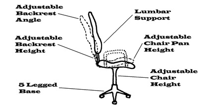
 Figure 1: A good chair should have several adjustable features.


  1. Lifting:

            When lifting the common adage of ‘lift with your legs, not your back’ is a myth! This has been propagated since the 1930s16;17 and has not reduced the incidence in low back injuries.18 In fact, many have questioned the validity of such a thing as a single universal lifting technique.17;19;20 This is because above all else the goal to lifting should be to: 1) Reduce the reaction moment (ie. by carrying objects close to the body) which reduces the extensor forces and the resultant compressive low back loading; 2) Avoid a fully flexed posture (ie. maintain the neutral lordosis by bending at the hips) which minimizes low back shear loading.21 One method to maintain the lordosis while picking up light objects is to perform the golfer’s lift.  This involves bending the neutral torso over a single hip to reach down while balancing the body with the other leg out behind. It should now be apparent that by satisfying these two criteria that many postures may be successfully used to accomplish a safe lift.     

  1. Appropriate Timing of Spinal Loads

            When considering the order of activity performance, one must appreciate that the spine has a loading memory.21;22  That is, prior activity modulates subsequent spine function.  The spine may be at significant risk of a destabilizing injury after either cyclic or sustained loading.23 This is due to the viscoelastic tissues of the spine. For example, after cyclic loading (ie. repeated lifting), laxity in the soft tissues causes a reduced protective muscular reflexive activity due to mechanoreceptor desensitization.23 Thus, a lifting activity should not be performed immediately following prolonged sitting or stooping and repetitive tasks. Instead the patient should recover spine stability (achieve redistribution of the nuclear material and recover ligament stiffness)24;25 by simply standing (which is a good rest from sitting),26 or consciously extend the spine (easily achieved by reaching for the ceiling).4 Walking for five minutes could also be recommended prior to lifting24 especially if preceded by prolonged vibration.27     

            The spine has a diurnal variation that affects its biomechanics and ultimately its injury mechanics. In fact, intervertebral disc-bending stresses are 300% and ligament stresses are 80% greater after rising from bed in the morning than later in the afternoon.28 This is why it may be a dangerous practice to perform early morning full range flexions of the spine as would be required of lifting and stooped postures. Snook et al. (1998)29 have demonstrated that simply avoiding full flexion early in the morning is a successful strategy for recovering LBP patients. After 30 minutes of upright posture, 54% of the daily disc height loss is achieved which reduces the potential risk of early morning back flexions, after 3 hours one loses about 80% of the total daily height loss!30

            It is a wise recommendation to avoid full flexion postures soon after rising including those that may be a part of a daily exercise routine especially for those undergoing rehabilitation. This is one concept that is critically important. I have seen on many occasions where a patient starts to respond to care, then one day confesses frustration that they are not getting any better or regressing. Upon questioning them I invariable discover that they are so motivated to get well they never skip their intensive early morning stretches – of course, excessively flexing their backs first thing in the morning and therefore damaging their disks! Tell your patients to stretch later in the day, at least after lunchtime. Taking garbage out to the curb the night before garbage day is another good example of using this principle. 


See the next issue of the AJCC for the continuation of the 5 key contemporary ergonomic concepts.


References---See AJCC Online for detailed list of references accompanying this article


10.  Andersson GBJ, Murphy RW, Ortengren R, Nachemson AL. The influence of backrest inclination and lumbar support on lumbar lordosis. Spine 1979; 4(1):52-58.

11.  Chaffin DB, Andersson GBJ. Occupational biomechanics. 2 ed. John Wiley & Sons, New York, 1991

12.  Cailliet R. Neck and arm pain. 3 ed. F.A. Davis Company, Philadelphia, 1991

13.  Andersson GBJ, Ortengren R. Lumbar disc pressure and myoelectric back muscle activity during sitting. III. Studies on a wheel chair. Scand J Rehab Med 1974; 6:122-127.

14.  Scalet EA. VDT health and safety: Issues and solutions. Ergosyst Associates, Lawrence, Kansas, 1987

15.  Carter JB, Banister EW. Musculoskeletal problems in VDT work: A review. Ergonomics 1994; 37(10):1623-1648.

16.  U.S.Preventive Services Task Force. Screening for risk of low back injury. Guide to clinical preventive services: An assessment of the effectiveness of 169 interventions. Williams & Wilkins, Baltimore, 1989, pp. 245-249.

17.  Parnianpour M, Bejjani FJ, Pavlidis L. Worker training: the fallacy of a single, correct lifting technique. Ergonomics 1987; 30(2):331-334.

18.  Brown JR. Lifting as an Industrial Hazard.  1972.  Labour Safety Council of Ontario, Ontario Department of Labour.

19.  Graveling RA, Simpson GC, Sims MT. Lift with your legs, not with your back: a realistic directive? Brown ID, Goldsmith R, Coombes K, et al., editors.  910-912. 1985. London, Taylor & Francis. Ninth Congress of the International Ergonomics Association.

20.  Chaffin DB, Park KS. A longitudinal study of low back pain as associated with occupational weight lifting factors. Am Ind Hygiene Assoc 1973; 34:513-525.

21.  McGill S. The biomechanics of low back injury: Implications on current practice in industry and the clinic. Journal of Biomechanics 1997; 30(5):465-475.

22.  McGill SM. Biomechanics of low back injury.  22-23. 1995.  XV International Society of Biomechanics. July 2-6, Finland.

23.  Solomonow M, Zhou BH, Baratta RV, Lu Y, Harris M. Biomechanics of increased exposure to lumbar injury caused by cyclic loading: Part I. Loss of reflexive muscular stabilization. Spine 1999; 24(23):2426-2434.

24.  McGill SM, Brown S. Creep response of the lumbar spine to prolonged full flexion. Clinical Biomechanics 1992; 7:43-46.

25.  Magnusson ML, Aleksiev AR, Spratt KF, Lakes RS, Pope MH. Hyperextension and spine height changes. Spine 1996; 21(22):2670-2675.

26.  Callaghan JP, McGill SM. Low back joint loading and kinematics during standing and unsupported sitting. Ergonomics 2001; 44(3):280-294.

27.  Pope MH, Magnusson M, Wilder DG. Low back pain and whole body vibration. In: Kumar S, ed. Biomechanics in ergonomics. Taylor & Francis, Philadelphia, PA, 1999, pp. 233-241.

28.  Adams MA, Dolan P, Hutton WC. Diurnal variations in the stresses on the lumbar spine. Spine 1987; 12(2):130-137.

29.  Snook SH, Webster BS, McGorry RW, Fogleman MT, McCann KB. The reduction of chronic nonspecific low back pain through the control of early morning lumbar flexion. Spine 1998; 23:2601-2607.

30.  Reilly T, Tyrrell A, Troup JDG. Circadian variation in human stature. Chronobiology International 1984; 1(2):121-126.


Sunday
Apr012012

The Link Aborted Fetuses Have to Autism

Thomas D. Pound, M.Ed.

Jennifer Brandon, DC

INTRODUCTION

In Vermont, Seth Mnookin recently discussed his recent work, “The Panic Virus:  A True Story of Medicine,”7 a book that concludes that the fear of vaccines is based primarily on myths.  With several of their officers, past and present, in attendance, Vermont’s Department of Health sponsored the talk. Mnookin’s logic follows the standard party line of the pharmaceutical industry; scientists, such as Andrew Wakefield, who lack any level of ethos, create the fear with false claims and biased research.  Influential advocates and celebrities, including Jenny McCarthy, then exacerbate the fear.  Mnookin’s bottom line?  Vaccines are beneficial.

On this particular evening, Mnookin did allow for a question and answer session.  Anecdotally, it appeared that out of the approximately 50 in attendance, only two were anti-vaccine.  Dr. Erin Sepic, a well-versed chiropractor from Richmond, questioned Mnookin’s claims that thimerosal was no longer a continuing factor as a risk effect of vaccines.  As it relates to autism, Mnookin’s answer during the talk was consistent with his position in his book where he writes about the “relative harmlessness”7 of the compound. 

Mnookin was then asked about recent studies indicating that the use of cells from aborted fetuses in the development of vaccines maybe accelerating the rate of autism.  He answered that he had not heard of that theory.  What is shocking is Mnookin’s lack of knowledge about an issue that is potentially bigger than the thimerosal controversy, because it has already reached the EPA and The Vatican.  It is alarming, because this is an issue where warnings were issued decades ago, and it is believed here that this will be the explanation for the rise in the numbers of autistic children.

Background

It is important to provide background.  Leonard Hayflick and his Wistar Institute developed a line of cells from an unborn fetus labeled WI-38.3  Additional fetal lines were also developed later4 for the purposes of developing vaccines for 31 separate viruses including those more familiar to the public such as measles, polio, varicella, rabies, herpes simples, and influenza.3  Currently vaccines for rubella, hepatitis A, and varicella were developed using the WI-38 fetal line.8  The MMR-II (introduced in 1989), the Varivax vaccine (introduced in 1995), and the VAQTA vaccine (introduced in 2002) all use the WI-38 fetal line for their respective virus propagation.11


DISCUSSION

It should not be surprising that those who are against abortion have already spoken out against these vaccines; even The Vatican issued a ruling on the matter.9 Previously, warnings about the use of fetal cells were theorized by no less an authority than Kevin McCarthy, the developer of the measles vaccine, when he stated one of two things he worried about “…in regard to WI-38 cell substrate,” and highlighting, “…the possibility of there being human genetic material passed over into the vaccine.2

It appears that Dr. McCarthy’s warnings should have been heeded.  Studies are now indicating that the years 19885 and 19951 are change point years, showing exact periods where the rate of autism accelerated.  If the theory holds, then 2002 should show itself as a change point year, since that was the first year for the introduction of the hepatitis A vaccines.  Ironically, Andrew Wakefield and his team predicted, “If there is a causal link between measles, mumps, and rubella vaccine and this syndrome (autism), a rising incidence might be anticipated after the introduction of this vaccine in the UK in 1988.”12


SUMMARY

It is unfortunate that this latest work in support of vaccines has left this important issue out of the discussion.  The possibility of vaccines having a genetic causal link to the rise in autism spectrum disorders is more than “tenuous”.6 As long as this theory is not part of any discussion about the harms from vaccines; it puts more children at risk with each injection.  The Vatican has spoken out against the use of fetal cells, the EPA has now looked into the issue, and the scientific community is now studying the link.  If Mr. Mnookin is going to hire himself out to speak in support of these poisons, then he needs to look into this issue deeper.


References

10.  Stephenson, R. W. (1948). Chiropractic Textbook. Davenport, Iowa: The Palmer School of Chiropractic.

11.  United States Food and Drug Administration. (2010, October updated 20). Vaccines Licensed for Immunization and Distribution in the US with Supporting Documents. Retrieved from http://www.fda.gov/BiologicsBloodVaccines/Vaccines/ApprovedProducts/ucm093839.htm

12.  Wakefield, A. J., Murch, S. H., Anthony, A., Linnell, J., Casson, D. M., Malik, M., et al. (1998). Ileal-lymphoid-nodular hyperplasia, non-specific colitis, and pervasive developmental disorder in children. The Lancet, 637-641.


Sunday
Apr012012

CBP Chiropractors: We Must Practice What We Teach


Cindy Boyd, BS, DC

Private Practice of Chiropractic Alameda, CA

Faculty Life Chiropractic College West

&


Deed E. Harrison, DC

President CBP Seminars, Inc.

Vice President CBP Non-Profit, Inc.

Chair PCCRP Guidelines

Editor—AJCC


INTRODUCTION

In previous issues of the AJCC, we have presented a number of articles on the indications and contra-indications of the cervical denneroll orthotic device as a method for restoration of an abnormal cervical lordosis. Also, several patient case studies have been presented describing the successful ability of the cervical denneroll orthotic device in restoring the cervical lordosis and improving patient health disorders as a consequence. In the current case, we report on the improvement in the cervical spine with a large disc herniation and complex injury-buckling of the cervical segments including retro-listhesis, hyper-extension, and flexion at different cervical spine joints. This report represents the actual conditions of one of the authors (CB) and these are her results.

Key Case Features

A 34 year old female presented with chronic neck pain, decreased range of motion, headaches with extension of the head and neck and a recent onset of heart palpitations of unknown etiology. The symptoms were reportedly getting worse over the past few months, and causing the patient to refrain from practicing in her chiropractic clinic providing one on one patient care. After administering 1-2 adjustments on any given day, the symptoms were exacerbated causing disability and significant discomfort.

The patient had a past history of two head and neck injuries. The first direct impact injury was sustained during a bicycle accident that occurred in the spring of 2008. The patient was riding a bicycle at a high speed and traversed over old railroad tracks at an awkward angle causing an immediate crash. Her head hit the pavement with significant force, and unfortunately she was not wearing a helmet. Fractures were ruled out and no lacerations were present. A mild concussion was sustained. Bruising and swelling was present in the right zygomatic region at the site of direct impact. 

Two years later, a double direct impact injury occurred during a snowboarding accident where the patient fell on an ice patch directly landing on the sacrum and a second impact occurred during the same fall when she fell backwards and hit her skull on the ice patch with significant force. This time the patient was wearing a helmet.  

  • ·      Health Status, Pain, and Disability Questionnaires

The short form (Sf)-36 health status questionnaire, numerical rating scale for pain intensity and the neck disability questionnaires were administered. The patient had considerable pain, disability, and depressed health. See Figure 1 for the initial NDI and SF-36 abnormalities.

Figure 1. Patient initial neck disability and SF-36 scores indicating considerable pain, disability, and abnormal health status.

  • Radiographic Evaluation

Lateral Cervical Radiograph:

On 8/24/11 a digital lateral cervical was obtained. See Figure 2. The radiographic analysis was done using the Posture RayÒ computerized radiographic mensuration system. The patient’s radiographic displacement values are shown in Figure 2B and are compared against normal. Several cervical spine subluxation-displacements are present including: an abnormal kyphosis from C4-C7, a C3-C4 segmental retro-listhesis, and a considerable hyper-extension of C2-C3 were identified. The radiographic and clinical findings indicated a possibility of herniation one or more cervical discs and instability in multiple levels of the cervical spine.  Thus, an MRI was obtained, and the findings confirmed a central/sub-ligamentous disc herniation at C2/C3 and C5/C6 with concomitant instability.

Figure 2. Initial lateral cervical radiograph and abnormal findings of alignment compared to ideal values. The PostureRay™ system was used.


Lateral Thoracic Radiograph:

            On 8/24/11 a lateral thoracic radiograph was also obtained. Figure 3 depicts the patient's lateral thoracic radiograph. Relative to the CBP ideal thoracic elliptical model, the patient's upper and mid-thoracic segments are in a relative subluxated extension position. This is consistent with an extension abnormal postural position of the thorax relative to the pelvis or lumbar spine.

Figure 3. Lateral thoracic radiograph. The green elliptical line is the Harrison ideal thoracic model representing the proper path of the posterior vertebral bodies of the thoracic spine. Note that the patient has extension-backwards bending of the upper-mid thoracic spine relative to the lower thoracic spine.

Denneroll Stress Radiography:

These values and findings indicate the patient’s candidacy for the Denneroll Cervical Orthotic. Based on MRI findings and significant abnormal segmental cervical translation measurements, Denneroll stress X-rays were taken using the large cervical Denneroll. Figure 4 depicts the lateral stress x-ray. Based on the initial radiographs, it was determined additional upper thoracic flexion and translation was needed in this setup to make a considerable correction in the injured and postural distorted regions. Thus, a 10 millimeter foam block was used under the denneroll device to increase the height of the orthotic in order to induce additional upper thoracic flexion and translation during the stress study.  The peak of the denneroll was placed in the lower cervical spine. Particular attention was given to the hyperextended upper cervical segments, and another block measuring 10 millimeters in height was placed under base of the skull during the stress analysis. Further, to limit the amount of skull extension and upper cervical extension, the patient was asked to actively flex-tuck her chin (although painful) at the time the radiograph was taken. Figure 2 shows the upper cervical spine extension on the initial patient x-ray. 

The stress analysis study showed considerable correction in the cervical abnormal values including the segmental translations and relative rotation angles that were present on the neutral lateral cervical study.

Figure 4. Denneroll stress lateral thoracic radiograph to ensure proper location of the denneroll peak and if the denneroll is effectively improving the cervical alignment.

  • Denneroll Cervical Orthotic Intervention

Based on the outcomes of the stress radiographic analysis, the patient agreed to participate in a study to determine the outcome of cervical curve correction using the Denneroll home traction device.  No other forms of treatment were administered. Spinal manipulation, CBPÒ drop table adjustments, Mirror ImageÒ exercises and in office traction types, were all avoided during this particular patient treatment phase with the Denneroll. 

The large cervical Denneroll was used at a frequency of 1-2 times daily, 4-6 days a week for a 30 day period. Each home session involved the patient lying supine on the large cervical Denneroll with a 10 millimeter block under the device. Refer to Figure 4 for the denneroll setup. In addition, a 10 millimeter block was placed under the skull to limit the amount of upper cervical extension.  The patient was also instructed to actively flex the chin while on the device. The amount of upper cervical hyperextension and thoracic extension measured on the lateral cervical and thoracic radiographs warranted the degree of specificity in this setup. Between the dates of 8/24/11-9/22/11 approximately 36 home Denneroll sessions were performed at a duration of 10-18 minutes each.

A follow up lateral cervical x-ray was taken on 9/22/11 to determine if the intervention had made any significant changes.

  • Case Outcome

      Subjectively, at the end of the 1-month treatment phase, the patient was asymptomatic including the disappearance of the heart palpitations, headaches and pain.  There were significant improvements in the SF-36 and neck disability outcomes. The follow up lateral cervical radiograph exam showed significant improvements in all cervical regions that were abnormal initial. Figure 5 and Table 1 describe these results.

Figure 5. One-month follow-up lateral cervical. Note that this x-ray was taken a minimum of one day with no treatment interventions. There are considerable improvements in all radiographic abnormalities.

RRA’s

(-) values = extension

(+) = flexion

Segmental Translation

(-) values = posterior

Translation C2-C7

C2/C3

12°

-1.6 mm

21.6mm

C3/C4

-1.7°

-2.4mm


C5/C6

-0.8 mm


ARA C2-C7

-26°


CONCLUSION

            This case report represents the actual conditions of one of the current study authors (C.B.). The uniqueness of this case lies in the severity of the cervical spinal displacements and the concomitant improvements in structural and functional abnormalities using the cervical Denneroll as the exclusive treatment intervention. While the patient's follow-up radiograph is still not considered to be within normal limits and further care is warranted, the improvements over the course of one-month are promising. The patient (C.B.) has committed to continuing her Denneroll intervention program to continue rehabilitating her cervical spine. Afterall, as a CBP Chiropractor, we must practice for ourselves what we teach our patients to do.


Sunday
Apr012012

CBP® NonProfit, Inc. Research Update

Paul A. Oakley, M.Sc., DC

CBP Research & Instructor

Private Practice New Market, Ontario, Canada


CBP Non-profit Supports Research Into the Effects of Adjustments on the Sympathetic Nervous System


CBP Non-profit, Inc. has joined the Australian Spinal Research Foundation and the William M. Harris Family Foundation to support a research study to investigate the effects of chiropractic adjustments on the sympathetic nervous system. The study’s Principal Investigator, Christopher J. Colloca, D.C. formulated the research design to study the effects of cervical spine adjustments in an animal model of cervical disc degeneration while measuring needle electromyographic (nEMG), spinal nerve root, and neural responses from the sympathetic chain. The study began in December 2010, and data collection was recently completed in December 2011. Results will be forthcoming in scientific conference presentations and journal publications.

Dr. Colloca and his team have a proven track record of publications into the biomechanical and neurophysiological effects of adjustments and dynamic spinal stiffness assessment using the validated intervertebral disc model in sheep at the Adelaide Centre for Spinal Research in Australia.  This study adds to the line of investigation underway at this laboratory in conjunction with pathologist, Robert J. Moore, Ph.D., and Belgian spine surgeon, Robert Gunzburg, M.D., Ph.D. Dr. Gunzburg serves as the Assistant Editor in Chief of the European Spine Journal, and has been collaborating on research projects with Dr. Colloca for fourteen years.

Research into the benefits of chiropractic adjustments have traditionally focused upon the area of the musculoskeletal system where findings have substantiated chiropractic care in the management of many conditions including low back pain, neck pain, and headache. This international research collaboration (Australia, Belgium, USA) brings together investigators from the professions of chiropractic, orthopaedic medicine, and anatomy and pathology to study how chiropractic adjustments can influence nerves in the body that control physiologic processes, including organ function, blood pressure and heart rate among others. 

Using a validated animal model approved by a governmental ethics committee, measurements of nerve activity will be conducted during chiropractic adjustments delivered with a hand-held mechanical adjusting tool. In this manner, the study can determine the effect of chiropractic thrusts on nerve function to better understand the mechanisms responsible for the improvements that many patients experience with chiropractic care. This research will not only build on the body of scientific knowledge in this area, but will be one of the few studies using a “lesion” model to investigate the neurophysiologic effects as opposed to studying normal populations. 

The $20,000 annual grant provided by CBP Non-profit matched a two-year $20,000 grant by ASRF and the $25,000 matching grant that William M. Harris Family Foundation has provided to the International Spine Research (INSPIRE) Foundation where Colloca is a Director.  Membership in CBP Non-profit, Inc. can be obtained online by visiting www.idealspine.biz and browsing through to the Research section of the product store. Members receive voting privileges at CBP Non-profit meetings and a Research DVD containing all of the CBP supported research publications.