Entries in Chiropractic BioPhysics (23)

Sunday
Jan112015

Treating the Elderly with Chiropractic BioPhysics® or CBP® Technique Methods

Jason W. Haas, DC

Private Practice Windsor, CO

Many practitioners are surprised to find out the extent of Chiropractic BioPhysics or CBP Technique methods we employ to treat elderly patients in our facility.  Some colleagues feel that the elderly patients may not be a candidate for treatment due to the fact that they are older, frailer, can have extensive degeneration or many comorbidities.1-3 These doctors are afraid to attempt to change their spine and posture because the problem is long-standing or they fear they may injure the patient.  However, in our clinical experience treating over 6500 patients in the last eleven years, we have found that the elderly patients respond very well to gentle application of CBP methods and astute clinicians will find that they can have a tremendous positive effect on a patient’s posture, pain, quality of life and overall health.

                  Most of our elderly patients come to us for painful conditions that they have not been able to find relief from using traditional treatments of drugs, surgery or even traditional chiropractic and physical therapy.  These patients are often discouraged with healthcare in general because they are finding that the time they spend with their doctor is less and less and the outcomes of the medicines they are prescribed are less and less effective. This can create a situation of frustration that a well-trained receptionist must contend with in order to get the elderly person feeling comfortable in the office.  A great tour of the office and a compassionate staff can show these folks that the CBP experience will likely be completely different from other chiropractors, medical doctors and other healthcare providers

Our Intake Process & General Exam Procedures

                  Once they see that we offer a different approach to spinal care, we can move them through the process of intake.  The examination is similar to younger patients but may require more time due to a longer health history and multiple concomitant health concerns/conditions.  We will always perform a thorough health history, all pertinent orthopedic and neurological testing, blood analysis if necessary, CBP structural evaluation including spine and extremity films when indicated, Posture Screen™ digital postural analysis and all pertinent outcome measures (all patients receive the SF-36, pain and disability questionnaires), and digital range of motion and strength testing.

                  Once we have the data for our initial assessment we make a determination as to whether they are candidates for CBP Structural Rehabilitation; yes there are some cases that we won't perform CBP care with due to contra-indications.  This assessment involves looking at the evaluated spine films with PostureRay™, and determining if there are any areas that would not respond well to conservative care, and CBP care especially.  If there are areas of suspected instability we will take flexion/extension films or send to my friend Evan Katz,DC for DMX.  Once possible contraindications have been ruled out, we explain the importance of improving sagittal and coronal balance with the patient and discuss the limitations that the care may have.  The discussions on limitations are crucially important because the elderly patients must be aware that osteophytes, disc and ligament degeneration and severe structural abnormalities will likely not return to normal as a result of the care we are providing.  They must know that the goals are better range of motion and strength, better posture (coronal and sagittal) and improvements measured on their outcome measures (SF-36, NDI, etc.) If a patient has an unrealistic expectation of being “fixed” and getting back to completely normal, it’s important to inject some reality and sometimes limit expectations.

Sagittal Plane Alignment & Health Concerns in the Elderly

                  The sagittal plane alignment of the spine and posture and it's connection to human health and longevity is becoming one of the most widely researched topics in spine care today.4-10 The sagittal plane alignment of the spine and posture in the elderly has been found to correlate to the following health disorders:

  1. Increased risk of spinal compression fractures;4
  2. Increased low back pain and more severe pain;5
  3. Decreased mobility and increased risk of falls leading to fractures;6,7,8
  4. Increased risk of going into a care giving facility and not being able to take care of one's self in normal activities of daily living;9
  5. Increased knee or patellar femoral pain;10
  6. Increased disability and impairment due to pain;11
  7. Possible increase risk of early death compared to age match corhorts.12,13

For example, of a few of these important studies are reviewed here. Kobayashi and colleagues4 prospectively followed 100 subjects aged 61.9 yrs of age for an additional 12 years (indicating they were about 74 years at follow up). Full spine radiographs were ascertained at initial and long-term follow up in an attempt to identify if sagittal plane radiographic alignment variables play a role in the risk for developing new vertebral compression fractures. In both univariate and multivariate analysis, reductions in lumbar lordosis (Cobb L1-L5) and thoracic kyphosis (Cobb T4-T12) increased the relative risk of developing a new vertebral body compression fracture. Significantly, even curves one standard deviation below the mean value showed statistically significant increased relative risks (RR 3.06). Their4 most statistically significant model was multi-variate including pre-existing compression fractures with both the lumbar lordosis and thoracic kyphosis decreased (RR 8.64). Kobayashi and colleagues4 suggested that flattened curves reduce the shock absorbing capability of the sagittal curves, increasing the dynamic loads on the vertebral bodies thus increasing the risk of fractures.

                  In a prospective study of 253 chronic LBP patients matched by age and physical characteristics to 253 normal controls between the ages of 50-85 years, Tsuji et al5 found a reduced L1-S1 lordosis in the chronic LBP group. Of primary importance, lumbar lordosis was inversely correlated with pain intensity on a visual analog scale (p= 0.025). In other words, as the lumbar lordosis decreased, the pain intensity of the subject was increased.5

                  Recently, Kamitami et al9, studied the spinal posture in 804 participants (65–94 yrs of age) who were initially independent in their activities of daily living (ADLL) at baseline. These participants were followed for a 4.5-year follow-up period and it was found that 126 (15.7%) of the participants became dependent in their ADLs. Dependence in ADL was defined as admission to a nursing home or need of home assistance to perform basic self care functions. Importantly, inclination of the upper body relative to the pelvis (angle subtended between the vertical and a line joining C7 to the sacrum) was correlated with outcome and lumbar curvature also showed a marginal association. After adjusting for age and sex, it was found that for each 1 unit increase in the quartile of forward inclination that the odds of becoming dependent on ADL's was 1.79 x greater. Indicating that the highest quartile had a risk of 1.79 x 3 = 5.37 times more likely to be dependent. This study is very important for the elderly person wanting to remain able to perform basic care functions.

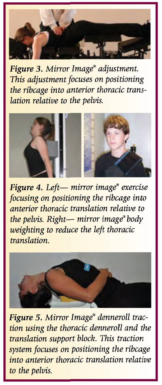
Chiropractic BioPhysics Mirror Image® Treatment Approach

                  Care of the elderly patient is slightly different in the CBP setting as it requires a gentler; yet still patient centered approach. We are cautious not to use high-velocity manipulation with elderly patients and work primarily on improving their posture with gentle CBP Mirror Image adjustments. The CBP Mirror image exercises are incredibly important in increasing strength and stability of the core postural muscles. We recommend and use the PowerPlate® as this is an exceptional tool for the elderly to perform their Mirror Image exercises while getting maximal benefit from the whole body vibration or acceleration training.

                  The CBP Mirror Imag traction protocols must also be altered for the elderly patients, they must understand why we are using the traction and stressing the analogy that spinal change is a marathon and not a sprint is important to minimize injury.  Most elderly patient in our office have radiculopathy, peripheral neuropathies, disc herniations and osteophytes so the use of distractive traction is often superior to compression traction which can irritate and worsen radiating symptoms.

                  Frequent re-evaluation and re-assessment of the elderly patient is possibly more important than any other age group.  This is due to the fact that they often have significant health complications that ned to be assessed periodically.  In our integrated setting it is easy for them to see our nurse practitioner or medical doctor if new symptoms arise, and for those of you who are not integrated, I would suggest having a close relationship with local providers to manage any health complications that may arise throughout care.

                  Diligent, patient, and compassionate care for the Elderly population can provide this segment with significant health gains, better posture and balance, less pain and a better overall quality of life. IF you would like more information on CBP care and the elderly, please attend any upcoming seminar.

Sample Patient Case:

Age: 87

Initial Complaints:  Low back pain and left leg sciatic pain NRS 8/10

Oswestry:  58% disability. Significant depression due to worsening of pain, poor balance and coordination.

Patient was told by two other chiropractors they could not help her.  She was told by a surgeon that she is not a candidate for surgery due to advanced age.

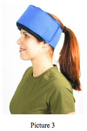
CBP Treatment:  Mirror Image adjustments prone with the headpiece elevated and instrument adjustment to the spine. Mirror Image exercises; standing with a block in the lower thorax and head and ribcage retraction performed in-office and at home.  Supine thoraco lumbar traction. Fulcrum of traction at thoraco-lumbar junction, 0° angle of pull and leg strapped below the femur heads, sustained for 12 minutes. Cervical Traction consisting of  Pope-2-way type traction with a distractive force of 10 lbs and a finishing front weight of 18 lbs. sustained for 10 minutes.

Total treatments:  30 sessions

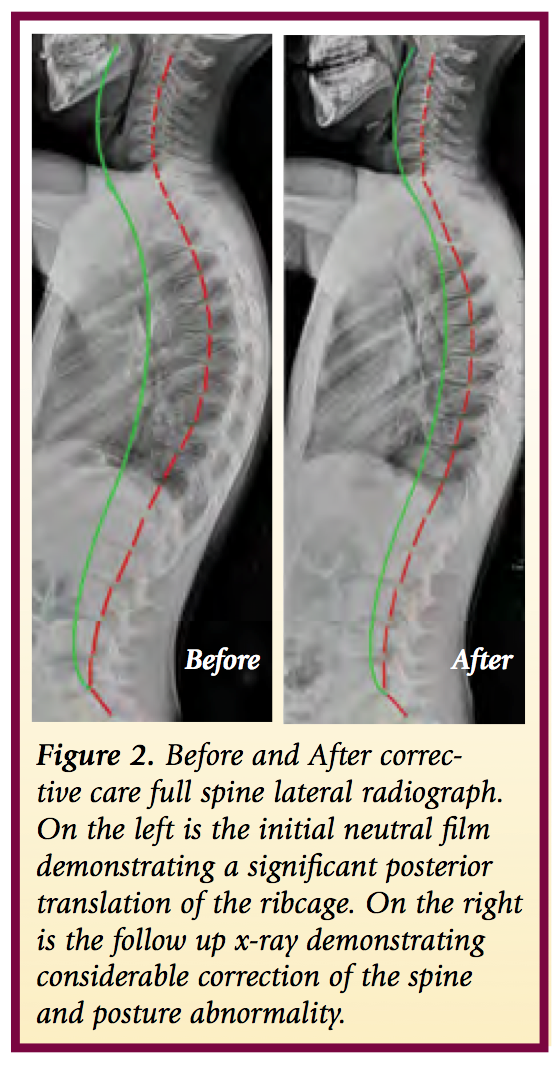
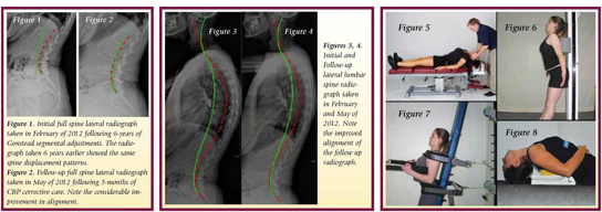
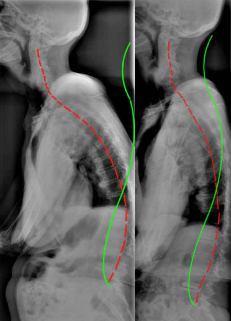
Figure 1. Before treatment and after treatment full spine radiographic changes due to CBP mirror image care over the course of 30 visits. Note that the follow up x-rays are taken a minimum of 3 days after the patients last treatment session. Thus, this is not an x-ray immediately after care. In this manner an accurate response to care can be found.

Final Complaints:  Very rare low back pain (NRS2/10)  Sciatica resolved, Depression significantly reduced and the patient states she feels better now than she has in decades. Oswestry: 8%.

Perspective on Patient Outcome:

                  It is my opinion that the improvement in the patient's condition, outcome measures, and self reported ability to function was due to the considerable improvement in the sagittal plane alignment of the patient's thoraco-lumbar curvatures and sagittal-forward balance. The references4-13 provided below provide evidence based support for this anecdotal but clinically obvious statement.


Reference Links

  1. http://www.ncbi.nlm.nih.gov/pubmed/25436061
  2. http://www.ncbi.nlm.nih.gov/pubmed/23307577
  3. http://www.ncbi.nlm.nih.gov/pubmed/25533322
  4. http://www.ncbi.nlm.nih.gov/pubmed/?term=Kobayashi+T+Osteoporos+Int++2008
  5. http://www.ncbi.nlm.nih.gov/pubmed/11479757
  6. http://www.ncbi.nlm.nih.gov/pubmed/21198460
  7. http://www.ncbi.nlm.nih.gov/pubmed/24715607
  8. http://www.ncbi.nlm.nih.gov/pubmed/20480146
  9. http://www.ncbi.nlm.nih.gov/pubmed/?term=Kamitami+2013
  10. http://www.ncbi.nlm.nih.gov/pubmed/12355123
  11. http://www.ncbi.nlm.nih.gov/pubmed/15972617
  12. http://www.ncbi.nlm.nih.gov/pubmed/19451575
  13. http://www.ncbi.nlm.nih.gov/pubmed/15450042
Sunday
Jan202013

3-Point Bending Traction for Scoliotic Curvatures Using the New 3-D Denneroll Traction System: A Case Report

INTRODUCTION

In a study from 1893 regarding scoliosis treatment, Bradford and Brackett,1, stated, “there is not only nothing irrational in the method of treatment by forcible mechanical correction when feasible, but it is manifest that when shortened ligaments in spinal curvatures are situated so that they serve as a check to muscular action.”1 They continue, “when they [ligaments] are strong enough to withstand muscular action, gymnastics [exercises] alone are inadequate as a system of correction.” 1 Bradford and Brackett’s1 mechanical traction protocol required patients to undergo traction for a half-hour daily. Because this study was done prior to the invention of x-ray, reported results were not very accurate. More than a century later, CBP® researchers and clinicians have found agreement with Bradford and Brackett that exercises should be combined with short duration, high-force mechanical traction in order to obtain the most effective results in scoliosis reduction.

  • CBP's Mirror Image® Traction for Scoliosis

The traction employed by the CBP® practitioner for scoliosis management requires critical reasoning and a thorough understanding of the displacements of the spine and posture. Generally speaking this traction is of the 3-point-bending type of load application or a transverse load applied at the apex of the curve with and without lateral bending, axial rotation, or other movements depending on the specific case. The traction set-up must always be performed in a pre-determined optimum sequence of movements using stress x-rays to guide the decision making process. Mirror Image® traction sessions and duration should be a minimum of:

  • At least 3-5 times per week. If the patient will traction more than 1 time per day this would be beneficial as long as the patient is not becoming overly painful from the increased frequency of treatment.
  • Traction duration should be 20-30 minutes. The patient starts with 2-3 minutes and over consecutive sessions progresses in time.

CASE REPORT       

            The current patient had a history of thoracic pain and had been under chiropractic care for many years which she indicated gave her temporary relief. Now at 13 yrs old, her pain and frequency have worsened over the last 4 months to a stage where she was experiencing daily headaches and thoracic pain rated as severe on a numerical rating scale (7-8 / 10).

  • Initial Radiography
  1. Primary Right Thoracic curve = 43 degrees (see Figure 1).
  2. Secondary Left Lumbar Curve = 28 degrees (see Figure 1).
  • 1st in traction x-ray using the Denneroll Table and the Scoli-Roll Fulcrum System

            The first in-traction x-ray showed that the thoracic spine was well effected however the lumbar spine was bending and under the stress in the incorrect direction (see Figure 2). This showed us that we needed to raise the lumbar spine off the table to help stretch the lumbar spine correctly.

  • 2nd in traction x-ray

            In response to the first in-traction x-ray, we decided to raise the pelvis to a level of +2 (two blocks under the right hip to address the concerns of the lumbar spine translation. You will see in the 2nd in-traction x-ray that raising the pelvis height did not decrease the effects of the ScoliRoll under the thoracic spine. This is obviously achievable due to the downward pressure of the two straps pulling on the thoraco-lumbar spine and upper thoracic region. The specific effects of using the block system to raise the pelvis is really evident when you look at the stress x-ray in figure 3.

From these in-traction x-rays we can accurately assess that the block under the pelvis is best for the patient’s spine. It also shows how x-rays are essential in establishing the best possible traction position.

  • CHIROPRACTIC INTERVENTIONS

            Due to the positive findings of the stress radiographs, the patient was recommended to undergo corrective chiropractic care including Mirror Image traction on the denneroll table, Mirror image adjusting, and Mirror Image Exercises. She was seen for 3 x week for 1-month (with a couple of interruptions) and was advised on doing home exercises on the days she was not being treated in the office.

  •  Mirror Image® Exercises and Adjustments

            We believe that both postural based exercises and adjustments are vital in consolidating the benefits of the effective spine stretching using the denneroll 3-point bending traction table. During the patient's exercise, neurological stimulation was added by impulsing the spine during her exercise movements; thus turning the exercise into the adjustment.

After 5 weeks and 13 sessions, we can see the corrective improvements in the patient’s spine. The patient’s symptoms have been reduced 90%. Thus, she is symptomatically doing very well and began improving after her 1st session and has reported no symptoms at all for the last 3 weeks.

  • 5 weeks-Follow up Radiography

            A one month follow up radiographs of the thoracic and lumbar spines were obtained to identify if the recommended and applied treatment was having the desired effect. Obviously scoliosis of this magnitude might require more frequent and increased numbers of sessions. However, only a follow up radiograph can truly determine what extent more care or different care is required.

            A remarkable reduction of the AP Thoracic scoliotic curve was identified from 41 degrees down to 28 degrees on the post (a 13° net improvement). Similarly, the AP Lumbar curvature demonstrated improvement. See Figure 5.

SUMMARY

            This case presents the initial successful reduction of a primary thoracic scoliosis in an adolescent female with a history of chronic pain. After 5 weeks and 13 sessions, we can see the corrective improvements in the patient’s spine. The patient’s symptoms have been reduced 90%. We believe the results are due to the combined effect of the Mirror Image treatment methods including the 3-point bending traction employed using the 3-D Denneroll Traction Table. The patient is continuing care and perhaps a future article will address her response.

References

  1. Bradford EH, Brackett EG. Treatment of lateral curvature by mean of pressure correction. 1893.

Sunday
Jan202013

Kinesiology Tape for Postural Control

INTRODUCTION

            As chiropractors, sometimes our efforts become frustrating if our careful attention to adjusting, stretching and strengthening, is undermined by patients resuming the same postural flaws that landed them in our offices in the first place. How exciting would it be to have a sticky, stretchy little assistant that reminded our patients for 2-5 days about positional awareness? Enter elastic therapeutic tape!

            By now, the vast majority of practitioners have had some exposure to elastic therapeutic tape or “kinesio-tape”, the commonly used brand name of developer Kenzo Kase. Tape companies claim it “reduces muscle soreness, improves function, decreases bruising, and decreases pain” and to some extent, these claims appear to be accurate.

            Anything that touches our body’s biggest organ, the skin, has a cutaneous mechanoreceptor effect that stimulates receptors to enhance body kinesthesia or movement awareness. By stimulating large skin mechanoreceptors, kinesiology tape can also downgrade painful stimuli from the nociceptors, which decreases pain perception.

            Early and persistent reasoning suggested that using the tape in an “origin to insertion”, or “muscle action” methodology, best serves to support/stimulate external body areas. While this approach probably makes the most intuitive sense to medical practitioners as it follows anatomical “rules of engagement”, emergent theories, which consider entire postural muscle groups, are making a strong case.

            Dr. Steven Capobianco, chiropractor and developer of the Fascial Movement Taping (FMT) method argues, kinesiology taping should be “based on the obvious yet largely overlooked concept of muscles acting as a chain… the body’s integration of movement via multi-muscle contractions as a means of connecting the brain to the body’s uninterrupted fascial web in order to enhance rehab and athletic performance via cutaneous (skin) stimulation. By taping movement rather than muscles, FMT has demonstrated greater improvement in both patient care and sport performance.

            Dr. Capobianco is not alone in this line of thinking. Leading fascia researcher, Robert Schleip PhD, underscores movement and its role in pain and dysfunction1. Additional support for this model comes from Thomas Myers in his ground-breaking book, “Anatomy Trains”2. He offers a template to assess, treat, and manage body-wide motor dysfunction based on myofascial meridians, and movement impairment.  

            Physical Therapy professor, Heather Murray, makes a strong case for the use of elastic therapeutic tape in those who maintain abnormal postures of the head and neck (i.e. in the work place). Her team conducted a pilot study3, which seemed to suggest that taping for scapular protraction could maintain better posture and decrease perception of pain.

            Recent research indicates that kinesiology tape has a greater stimulatory effect with compromised tissue (due to injury or fatigue due to poor posture). Thedon, et al4 conducted a study to evaluate body sway in individuals with and without tape. They found that the tape showed very little change in the uncompromised condition, but when the subjects were fatigued, the tape provided an added stimulatory effect to the skin helping to compensate for the loss of information fed to the brain from the muscles and joints. For the pain and performance community, this study provides insight into an “auxiliary” system, such as the skin, to augment treatment and training outcomes.

            A 2012 study5 of 32 surgeons, showed a statistically significant reduction in neck and low back pain (using Oswestry Low Back Disability Index and Neck Disability Index) and functional performance (using neck and low back range of motion scores) with the use of kinesiology tape during surgery. This may have far-reaching implications for other jobs/activities where sustained positions result in musculoskeletal pain.

            Let’s look at a common complaint where chiropractors can utilize elastic therapeutic tape to enhance postural control (awareness). Kinesiology tape is commonly used in chiropractic offices to enhance scapular retraction, alleviating upper extremity discomfort caused by internally rotating shoulders (especially when the scapula is in a protracted position). The tape, according to Dr. Capobianco’s model, is applied in a functional manner to augment its therapeutic effect. Begin by addressing the neuro-myo-skeletal dysfunction associated with the protracted shoulder girdle (manipulation, myofascial release, movement re-patterning, etc.). Once the patient is able to appreciate an appropriate retracted/depressed scapula, apply the kinesiology tape (see inset) in a manner to, appropriately, stimulate the local receptors once the intended position is lost.

  • Step 1: Manually mobilize/manipulate the thoracic spine and shoulder girdle and associated myofascial chains
  • Step 2: Place patient/athlete into appropriate postural position that centrates the scapula-thoracic segment.
  • Step 3: Tape the local area (see X pattern and H pattern) with “NO” stretch to stimulate local receptors
  • Step 4: Corrective exercises that will help with postural re-education.


SUMMARY  

            Clearly the use of kinesiology tape is popular (millions of users) and the applications are broad (from athletic injuries to edema). Specific evidence for efficacy is scant but growing, and plausible. There are currently no reported dangers associated with using this elastic cotton mesh bandage, and the only significant contraindication is on open wounds. Kinesiology tape breathes well and flexes like a second skin, unlike most braces that act more like abrasive exoskeletons. It withstands sweat and/or water and is by most comparisons a cost-effective treatment modality. While science is unlikely to discover that kinesiology tape is the panacea for all aches, injuries and postural distortions, medical practitioners should keep this tool in the chest due to its vast possibilities in treating patient complaints and as a tool for postural re-education.

References:

1. Schleip R, Muller D. Training principles for fascial connective tissues: Scientific foundation and suggested practival applications. J Body Move Ther 2012;1-13.

2. Myers, T.W. 2009. Anatomy Trains: Myofascial Meridans for Manual and Movement Therapists. New York: Churchill-Livingston.

  1. Effects of Kinesio Taping on Posture and Presence of Upper Extremity Pain

4. Thedon T, et al. Degraded postural performance after muscle fatigue compensated by skin stimulation. Gait Posture, 2011 Apr;33(4) 686–9.


Sunday
Jan202013

Combining Specific Chiropractic Adjusting Techniques with CBP Corrective Care Techniques: Case #2.

INTRODUCTION

            In our previous article (AJCC October 2012)1, we suggested that the astute Chiropractor should become skilled at administering both a segmental specific adjustment technique (such as the Gonstead system) and a full spine corrective technique like Chiropractic BioPhysics (CBP). In this manner, patients will experience the benefits of segmental motion restriction improvement and the restoration of proper full body and spine alignment. The case presented herein attempts to provide further rationale for the combination of these two uniquely distinct, but complimentary full spine Chiropractic Technique systems.

CBP / Gonstead Case Study #2:

            In 2012, an 8 year old male, who had no overt symptom complaints was brought to the author's (T.P) Chiropractic clinic for postural evaluation and a spinal checkup. His parents had noticed significant posture abnormality and were concerned about underlying spinal disorders.

  • Postural Analysis: Posture analysis revealed a significant left thoracic translation, a right lateral flexion of the ribcage, a right posterior pelvic rotation, a suspected left leg length inequality, a large posterior thoracic translation, and a slight forward head translation. 
  • Radiographic Analysis:  
  1. In April of 2012, a modified AP femur head-ferguson view was obtained. On the left of Figure 1, this x-ray is shown. The PostureRay x-ray software system was used to identify and quantify the extent of leg length inequality. A 9mm left leg length inequality was identified with consequent spine abnormality.

2. In April of 2012, a full spine lateral radiograph was obtained. While in June of 2012, a follow up full spine lateral was obtained to document the response to intervention and potential modifications that might be warranted. Figure 2 depicts these full spine radiographs

  • Interventions: A total of 24 treatment sessions over the course of 2 months was utilized in this case.

            For segmental adjusting technique, the treating Chiropractor (TP) exclusively adhered to and utilized the Gonstead Technique system for identified spine subluxations including:

  1. Abnormal temperature differential patterns (nervo-scope / tempo-scope).
  2. Static palpation data indicating the presence of edematous tissue around the injured segment.
  3. A decrease in motion of the segment in question, as compared to the surrounding area.
  4. Palpable muscle spasm or splinting around the area in question.
  5. Visualization of the area (looking for presence of pitting edema, asymmetry in the tissues, etc).
  6. Then, consulting the 3-shot, digitally stitched, AP full spine x-ray and the lateral (2 shot) full spine x-ray to analyze the “foundation principle” and relate this to the examination findings.

            For the full spine and posture corrective care, CBP® mirror image® adjusting, exercise, traction procedures were utilized. Additionally, body weighting using the thoracic belt from Circular Traction was applied 5 times per week for 15 min walking intervals. These procedures were administered on each visit to the patient. Lastly, a full foot lift of 9 mm was administered to the patient and was placed in the left shoe.

  • Mirror Image Adjustments

The patient was administered mirror image adjustments to correct posterior thoracic translation with hyper-kyphosis and anterior head translation with head flexion postures. See Figure 3.

  • Mirror Image Exercises

The patient was administered mirror image® exercises to correct posterior thoracic translation with hyper-kyphosis and anterior head translation postures. See Figure 4.

  • Mirror Image® Body Weighting and Denneroll Traction

      Mirror Image traction care was administered to the patient. The patient received the denneroll traction system to correct the abnormal thoracic translation posture as shown in Figure 5. In addition the patient was instructed in the use of the denneroll thoracic orthotic and was advised to do this 5 times per week at home.

  • Case Outcome

            Subjectively, at the end of 2 months of corrective care, the patient's posture was remarkably improved. NRS = 1-2 / 10. According to the patient's parents, in their own words, "It’s great to see how quickly he has improved. The leveling of his shoulders and hips is apparent and how he carries himself; he is dramatically more upright and balanced".

            Objectively, the patient's follow up full spine radiographs in Figure 2 show remarkable reduction of the spinal displacements.


CONCLUSION

            The authors' opinion is that the patient's improvements were directly related to both the segmental adjusting and spinal corrective procedures applied using CBP Technique. Accordingly, for optimum patient response, traditional Chiropractic adjustments would seem to be enhanced by the addition of spinal corrective procedures as in CBP. In the end, it is the positive response of the patient that should dictate this combined approach of classical Chiropractic care, with more contemporary corrective Chiropractic systems.

References

  1. http://www.chiropractic-biophysics.com/clinical_chiropractic/2012/12/18/combining-specific-chiropractic-adjusting-techniques-with-cb.html


Sunday
Jan202013

CBP® NonProfit, Inc. Research Update

Check Out Our Research Reference List Online at: http://www.idealspine.com/cbp-research/

Paul A. Oakley, M.Sc., DC

CBP Research & Instructor

Private Practice New Market, Ontario, Canada


            Chiropractic Biophysics Non-profit, Inc. is a 501(c)(3) nonprofit corporation dedicated to the advancement of chiropractic principles through scientific research. Dr. Don Harrison (deceased) and his second wife Dr. Deanne LJ Harrison (deceased) founded CBP research foundation in 1982; it was registered as CBP Non-Profit, Inc. in 1989 by Dr. Sang Harrison (Don’s 3rd and final life’s love). Through this organization Dr. Don and colleagues have published 140 peer-reviewed spine and Chiropractic research publications. Further, CBP Non-Profit, Inc. has funded many scholarships as well as donated chiropractic equipment to many chiropractic colleges; always trying to support chiropractic advancement and education. Dr. Don Harrison was the acting president of CBP Non-Profit, Inc. since 1982. Currently, Dr. Deed Harrison (Don’s son) is the President of CBP Non-Profit, Inc.

            Results of our studies have been published in prestigious research journals and presented at respected conferences around the world. Your (Chiropractic donations) support enables us to continue important research and gives you a voice in the course our studies take. Join today, either as a regular member or member of the President’s Circle. The result will be better chiropractic techniques, stronger chiropractic practices, and healthier chiropractic patients.

CBP® research consists of studies on a variety of CBP® technique related topics including:

  1. Spine modeling studies evaluating ideal and average human alignment variables,
  2. Spine biomechanics studies analyzing loads, stress, and strains,
  3. Posture modeling studies,
  4. Reliability of measurements and evaluation of patients (x-ray, posture),
  5. Validity of the measurements and evaluation of patients,
  6. Randomized trials evaluating technique outcomes
  7. Non-randomized trials evaluating technique outcomes,
  8. Case series studies evaluating technique outcomes,
  9. Case studies evaluating technique outcomes,
  10. Literature reviews and professional commentaries.
Tuesday
Dec182012

Colorful Olympic Tape for our Patients – TAPING MOVEMENT, NOT MUSCLES

Steven Capobianco D.C, DACRB, CCSP

Medical Director, Rock Tape.

www.rocktape.com


INTRODUCTION

            “Kinesiology tape” was first used by acupuncturists and chiropractors in Japan over 20 years ago. Today it is used by practitioners throughout the world in the treatment of injuries and to improve sports performance.

It was apparent that world-class athletes were seeking an alternative to managing pain and maximizing recovery potential at the 2012 London Olympic Games. Practitioners and spectators alike witnessed athletes, from the diving platform to track and field, wearing colorful tape in intricate patterns and with questions of what it was, word began to spread.. While the use of kinesiology tape is often associated with athletes, the reality is that kinesiology tape is effective for a wide variety of problems, not just sports injuries.

The evidence supporting the role of kinesiology tape still lacks high-level clinical trials, but if you spend the time to research further you will start to see a trend. This trend was spearheaded by Thibaud (Feb 2011), and reported the effects of taping the skin in subjects that are compromised, either fatigued or injured. They found that the skin acted as an auxiliary kinesthetic system providing the brain with postural awareness when the compromised muscle/joint receptors were fatigued.  

Kinesiology tape, developed and refined for the past 35 years, attempts to mimic the qualities of the skin in its elasticity and thickness. With these qualities in mind, the tape is applied on the skin in certain patterns to enhance certain physiological processes.  Once applied to the skin the elastic capacity of the tape  provides a light shear augmenting the mechanoreception of the area addressed. This  same elastic recoil aids in pulling the epidermis and dermis layers of skin away from the underlying fascial compartments, thus improving fluid dynamics (acute/chronic edema) (Ya-Hui Chou). Both of the above processes have a substantial benefit in down-regulating nociception (pain), in return improving movement patterns in accordance to the area addressed with tape. Lastly the tape in its ability to enhance kinesthetic awareness through skin stimulation, can improve postural dysfunction. Careful application of tape along “fascial strain lines” has been shown to improve common postural dysfunctions such as upper and lower cross syndromes, hyper and hypolordosis, anterior/posterior tiling of the pelvis, and more (see figure 1).

SUMMARY

The refinement of kinesiology taping has extended from the traditional anatomical (muscle) applications to the more integrated fascial (movement based) applications. This innovative way of taping has revised some new and improved taping applications that have shown to improve movement dynamics, such as postural sway associated with compromised environment like pain and fatigue states, as discussed earlier in the science of taping. Athletes, such as those in the Olympic Games, are interested in the advancement of taping technology. With this added interest in “legal” performance enhancement which doesn’t involve drugs, athletes and coaches are looking for an edge in other alternatives including kinesiology tape. By enhancing our body awareness via movement taping, we can improve the length-tension relationships of the associated muscles, and, in turn, improve the neuromuscular efficiency of the intended activity.

Recently, the Garmin-Barracuda professional cycling team has implemented this model of taping to improve postural position on the bike to optimize power outputs during long cycling time trials. The team’s physicians believe that the tape, applied in specific patterns, improves form which translates to a faster time with less incidence of injury. This all translates into improved performance without the use of illegal enhancements. Future studies on the effects of stimulating the largest organ in our body (the skin) via kinesiology tape, is a new direction to investigate with regards to improving proprioception, posture and performance.

References


  1. Fuller, R.B. 1975. Synergetics. New York: Macmillan
  2. Ya-Hi Choi, et al. Manual Lymphatic Drainage and Kinesio Taping in the Secondary Malignant Breast Cancer-Related Lymphedema in an Arm With Arteriovenous Fistula for Hemodialysis. American J. of Hospice and Palliative Med. 00 (0) 1-4, (2012).
  3. Fascia Congress. 2009. www.fasciacongress.org/2009
  4. yers, T.W. 2009. Anatomy Trains: Myofascial Meridans for Manual and Movement Therapists.  New York: Churchill-Livingston.
  5. Thibaud, et al.Degraded postural performance after muscle fatigue can be compensated by skin stimulation. Gait and Posture 33 (2011) 686-689.
  6. Hyun Mo An, et al. The effects of kinesio tape on lower extremity functional movement screen scores. International J. of Exercise Science 5 (3): 196-204 (2012). 


Tuesday
Dec182012

Combining Specific Chiropractic Adjusting Techniques with CBP Corrective Care Techniques

Todd Pickman, DC

Private Practice of Chiropractic Eagle, ID

Gonstead Technique & CBP Trained Chirorpactor


Deed E. Harrison, DC

President CBP Seminars, Inc.

Vice President CBP Non-Profit, Inc.

Chair PCCRP Guidelines

Editor—AJCC 


 INTRODUCTION

            We've all heard the Chiropractic adage that 'all techniques work'. This is likely true but we first need to define what is meant by the term 'works'. If we assume that what is meant by 'works' is that all techniques are able to improve joint mobility, posture mobility, and reduce pain and muscle hyper-activity by reflexo-genic inhibitor effects, then this statement holds some merit. However, certain specific segmental adjusting techniques have more research validating this statement than others. For example, using the Gonstead Technique system of analysis and adjusting, Nansel and colleagues1 found statistically significant improvements in cervical lateral bending motion responses following one specific adjustment to the restricted side of the cervical spine.

            In contrast, if we assume that what is meant by 'works' is that all techniques are able to improve the alignment of generalized abnormal sagittal plane curvatures, then this statement does not hold merit. For example, using the same Gonstead Technique system of analysis and adjusting, Plaugher et al2, found no measurable change in the cervical and lumbar lordosis in 50 patients receiving several adjustments. Regarding CBP Technique corrective methods, statistically significant improvements in sagittal plane alignments have been identified in several clinical control trials examining patients suffering from chronic low back and cervical spine disorders.3-6

            So what does the above information indicate? In our opinion, the above indicates that the astute Chiropractor should become skilled at administering both a segmental specific adjustment technique and a full spine corrective technique like Chiropractic BioPhysics (CBP). In this manner, patients will experience the benefits of segmental motion restriction improvement and the restoration of proper full body and spine alignment. The case presented herein attempts to provide initial rationale for the combination of two uniquely distinct, but complimentary full spine Chiropractic Technique systems.

Case Study:

            Approximately, 6. 5 years ago (in 2006), a 74 year old female presented to one of the author's (T.P) practices seeking care for the relief of functional disabilities related to a chronic low back condition. At this time, the Chiropractic clinician (T.P) exclusively adhered to and utilized the Gonstead Technique system of analysis and adjusting for identified spine subluxations including:

  1. Abnormal temperature differential patterns (nervo-scope / tempo-scope).
  2. Static palpation data indicating the presence of edematous tissue around the injured segment.
  3. A decrease in motion of the segment in question, as compared to the surrounding area.
  4. Palpable muscle spasm or splinting around the area in question.
  5. Visualization of the area (looking for presence of pitting edema, asymmetry in the tissues, etc).
  6. Then, consulting the 3-shot, digitally stitched, AP full spine x-ray and the lateral (2 shot) full spine x-ray to analyze the “foundation principle” and relate this to the examination findings.

            At this time, the patient was recommended and consented to a program of care consisting of Gonstead adjustments at a frequency of 3 times for 2-weeks, 2 x per week for 4 weeks, and then 1 x per week for 6 weeks. At this point the patient was improved to some extent (not completely) and was placed on a Chiropractic maintenance schedule consisting of 1-4 x per month sessions of adjustments over the course of 6 years. Through the course of this 6-year time period the patient would have episodic acute flare-ups of her chronic low back conditions that would improve with Chiropractic. The patient averaged a minimum pain intensity of 4/10 over the course of these 6-years with flare-ups reaching 8/10 on a numerical rating scale.

  • Feb 2012 Findings: In February of 2012, the Chiropractor (T.P.) informed the patient that he had learned a new approach to full spine corrective Chiropractic (CBP Technique) and that he believed she was a candidate for this 'new' system. The patient was recommended and consented to a 36 visits, 3-month, corrective care program consisting of the combination of Gonstead Technique (segmental adjusting applied to the cervical, thoracic, and lumbo-pelvic regions) and CBP Technique. Her pain scale was a 7/10, she reported difficulty walking more than 1/4 mile distances without severe thoraco-lumbar pain, fatigue, and spasm, all while wearing a back brace. 
  • Radiographic Findings:

      Lateral Full Spine Radiograph:  In February, 2012 a new digital full spine lateral radiograph of the patient was obtained. See Figure 1. The radiographic analysis was done using the PostureRayÒ computerized radiographic measurement system. Qualitatively, the patient’s radiographic displacements are shown in Figure 1 as the path of their posterior vertebral body margins from C2-S1 relative to an idealized spine model in upright stance developed by Harrison and colleagues.7 The patients follow up full spine lateral radiograph after 3-months of CBP Technique is shown Figure 2. Note the remarkable improvement in alignment, whereas previously, for 6 years no such improvement was identified with Gonstead Technique alone.

      Lateral Lumbar Spine Radiograph:  In February, 2012 a digital lateral lumbar radiograph was obtained. See Figure 3. The radiographic analysis was done using the Posture RayÒ computerized radiographic mensuration system. The patient’s radiographic displacement values are shown in Figure 3 and are compared against normal.7 The patients follow up full spine lateral radiograph after 3-months of CBP Technique is Figure 4. Note the remarkable improvement in alignment where for 6 years no such improvement was identified with Gonstead Technique alone.

Interventions: The primary course of interventions included CBP mirror image® adjustments, exercises, and traction to reduce her full spine postural and spine displacements. The treatment frequency was 3 times per week for 40 visits over approximately 12 weeks. The Patient presented to and actively participated at all appointments. Each visit consisted of mirror image adjusting, mirror image exercises and traction to reduce her abnormal posture displacements.

  • Mirror Image Adjustments

The patient was administered mirror image adjustments to correct posterior thoracic translation with hyper-kyphosis and anterior head translation with head flexion postures. See Figures 5.

  • Mirror Image Exercises

The patient was administered mirror image adjustments to correct posterior thoracic translation with hyper-kyphosis and anterior head translation with head flexion postures. See Figures 6.

  • Mirror Image® Traction

      Mirror Image traction care was administered to the patient. The patient received traction in the standing posture as shown in Figure 6. In addition the patient was instructed in the use of the denneroll thoracic orthotic and was advised to do this 5-7 times per week at home. See Figure 7.

  •  Case Outcome Subjectively, at the end of the 1st month of corrective care, the patient was remarkably improved; NRS = 1-2 / 10. Her low back condition improved to where she was able to walk 3-miles without the use of a low back support and without debilitating pain. According to the patient, in her own words, "I do know that I'm experiencing a life change for the better...I'm singing the praise for the treatment (CBP Technique--added for clarity) and how great I feel".


CONCLUSION

            The authors' opinion is that the patient's improvement is directly related to the spinal corrective procedures applied using CBP Technique. Accordingly, for optimum patient response, traditional Chiropractic adjustments would seem to be enhanced by the addition of spinal corrective procedures as in CBP. In the end, it is the positive response of the patient that should dictate this combined approach of classical Chiropractic care, with more contemporary corrective Chiropractic systems.

References

  1. Nansel DD, Cremata E, Carlson J, Szlazak. J Manipulative Physiological Ther 1989;12:6:419-427.
  2. Plaugher G, Cremata EE, Phillips RB. J Manipulative and Physiol Ther 1990.
  3. Harrison DD, Jackson BL, Troyanovich SJ, Robertson GA, DeGeorge D, Barker WF. J Manipulative Physiol Ther 1994;17(7):454-464.
  4. Harrison DE, Cailliet R, Harrison DD, Janik TJ, Holland B.  Archives Phys Med Rehabil 2002; 83(4): 447-53.
  5. Harrison DE, Harrison DD, Betz J, Janik TJ, Holland B, Colloca C. J Manipulative Physiol Ther 2003; 26(3): 139-151.
  6. Harrison DE, Harrison DD, Cailliet R, Janik TJ, Holland B. Arch Phys Med Rehab 2002; 83(11): 1585-1591.
  7. Harrison DE, Harrson DD, Haas JW, Oakley P. Spinal Biomechancs for Clinicians, Vol I. Evanston, WY: Harrison CBP Seminars, Inc., 2003, ISBN 0-9721314-2-6.
Tuesday
Jul172012

American Journal of Clinical Chiropractic 

Tuesday
Jul172012

Lumbar Spine Ergonomics Part 2: The 5 Key Contemporary Concepts

Paul A. Oakley, M.Sc., DC

CBP Research & Instructor

Private Practice New Market, Ontario, Canada

VIEW PRINT VERSION


INTRODUCTION

     Last issue we discussed the essential reasons for the understanding of contemporary ergonomics concepts and their potential contradictory effects on patient care – good ergonomic practices will aid in patient response to care - poor ergonomic practices will undeniably compromise patient outcomes. We have discussed the first two concepts, and now continue with concept 3 of 5.

3) Optimal Spinal Loading: Not too Much, Not too Little

     A common ergonomic misconception is that recommendations should be directed at minimizing tissue loading, however, this is not true.1 For example, not typically considered stressful to the spine, sleeping for more than 8 hours at a time may indeed stress the spine.1 This is due to the fact that the discs swell by imbibing fluid over night,2 and since the discs function to transmit force, (rather than absorb force which is the function of the vertebral bodies)3 the column is subjected to increased stiffness and is at greater injury risk in the early morning.4 The fact is too much loading or too little loading is potentially injurious.  For this reason, “the challenge is to develop a wise break strategy to facilitate optimal tissue adaptation.”1


Since most spinal injury occurs as an accumulation of micro-trauma as opposed to an isolated acute traumatic event,5,6  regular ‘micro-breaks’ are recommended. This allows a continuously varying demand and subsequent migration of load on the low back tissues.1 Standing up to answer the telephone while performing seated work is a good way to aid in varying the posture throughout the day.  Any posture adopted for prolonged periods may fatigue the tissues used to maintain that position.  When standing for long periods one can alternate the placement of a foot on top a footrest to reduce tension in the psoas and lumbar spine.7 See Figure 1.   


     The idea of an ideal sitting posture is a farce. This is because it would only be ideal for about 10 minutes1 as sitting creeps the posterior ligamentous tissues (20 minutes of continuous sitting requires over 30 minutes of non-seated recovery time to regain the normal protective spine stiffness8). The ideal sitting posture is a variable one.9,10  McGill (2002)1 suggests three recommendations for prolonged sitting: 1) Use an ergonomic chair properly (i.e. vary the adjustable features regularly within sensible ranges); 2) Get out of the chair (i.e. rest breaks should involve opposite activities – Mirror Image Postures); 3) Perform an exercise routine during the workday (i.e. not first thing in the morning; not exercises that exert excessive spinal loads).

4. Reduce the Reaction Moment

The ‘reaction moment’ is the rotational force your body must generate to successfully perform a sagittal lift - the weight of the load (and upper limbs) would cause your torso to fall forward unless the back extensors offset this gravity-induced moment to allow the maintenance of an upright lift. The extensor activity within the low back causes compression of the spinal mechanism.11 Excessive compression within the low back is detrimental especially during prolonged, or repeated tasks. Excessive loading of the low back due to reaction moments can occur during any activity where there is a demand for the body to resist a force that is a distance from the spine.

Reducing the reaction moment is the key to reduce spinal compression. This is accomplished by carrying the load closer to the body and/or reducing the weight of the load lifted. See Figure 2.


Fig. 2: Reduction of ‘reaction moment’ and therefore muscle work is accomplished by bending the arm to decrease distance of arm segments and load from spinal axis.

     A third method of reducing a reaction moment is to direct the transmissible vector through the low back. The ‘transmissible vector’ is likened to the former concept only applied to tasks such as pushing and pulling. It should be known that there is no specific muscle that has the primary function of producing torso torque, it is performed by co-contraction of all the torso muscles resulting in substantial spinal loads.1 Thus, when performing tasks such as pushing and pulling on objects such as doors and vacuums, the pull/push should be directed so that it is in line with the low back.

5) Maintain Spine Stability

     McGill was the first to capture a ‘live’ segmental spinal buckling while viewing the spine of a power lifter during a lift.12 This buckling is possible when either there were high forces in the global muscles and low forces in segmental muscles or when there are low forces in all muscles.13 This mechanism explains how one can injure themselves by performing ‘negligible’ lift such as picking up a pencil.5

     As expressed by McGill, “stiffness creates stability,” and symmetrical stiffness offers greater stability.1 Thus, a slight abdominal co-contraction (5-10% max. vol. contraction) will ‘brace’ or stabilize the spine prior to a lift. This will prevent any tissue from bearing a surprise load.14 Spinal stability is also maintained by adopting symmetric postures, avoiding twisting action, and flexion postures. Symmetrical trunk postures also ensure greater available strength,15 enabling one to perform activities easier in symmetric postures.

     Lastly, the lumbar lordosis is essential in maintaining spinal stiffness – keeping it dynamically (lifting) and having it statically (standing) – any chiropractic treatment neglecting the correction of lumbar lordosis is missing a key ingredient for their patient’s back stability and health.

Conclusion

     It has been suggested that the “mechanics of the spine are not well understood by those who examine and treat these structures.”16 The Chiropractic BioPhysics® lumbar rehabilitation protocol has its unique niche in remolding the spinal posture and thus succeeding where most other LBP treatments fail including other chiropractic techniques. Both the neutral static posture and the dynamic postures performed throughout the day have significant implications for spinal health and response to treatment. The performance of a lumbar rehabilitation without regard for the dynamic activities performed throughout the day may render patient care fruitless. Always consider the Five Key Concepts to Lumbar Spine Ergonomics:

1. Maintain the Neutral Lumbar Curve

2. Appropriate Timing of Spinal Loads

3. Optimal Spinal Loading: Not too Much, Not too Little

4. Reduce the Reaction Moment

5. Maintain Spine Stability


References


1.McGill SM. Low back disorders: Evidence-based prevention and rehabilitation. Champaign, IL: Human Kinetics, 2002.

2.Urban JPG, McMullin JF. Swelling pressure of the lumbar intervertebral discs: Influence of age, spinal level, composition, and degeneration.  Spine 1988;13:179-187.

3.Ruch WJ. Atlas of common subluxations of the human spine and pelvis. New York: CRC Press, 1997.

4.Adams MA, Dolan P, Hutton WC. Diurnal variations in the stresses on the lumbar spine.  Spine 1987;12:130-137.

5.McGill S. The biomechanics of low back injury: implications on current practice in industry and the clinic.  Journal of Biomechanics 1997;30:465-475.

6.Kumar S. Cumulative load as a risk factor for back pain.  Spine 1990;15:1311-1316.

7.White III AA, Panjabi MM. Clinical biomechanics of the spine.  2 ed. New York: Lippincott Williams & Wilkins, 1990.

8.McGill SM, Brown S. Creep response of the lumbar spine to prolonged full flexion.  Clinical Biomechanics 1992;7:43-46.

9.Eklund J. Biomechanical aspects of work seating. In: Kumar S, ed. Biomechanics in Ergonomics. Philadelphia: Taylor & Francis, 1999:325-334.

10.  Pynt J, Higgs J, Mackey M. Seeking the optimal posture of the seated lumbar spine.  Physiotherapy Theory & Practice 2001;17:5-21.

11.  Troup JDG. Relation of lumbar spine disorders to heavy manual work and lifting.  Lancet 1965;857-861.

12.  Cholewicki J, McGill SM. Lumbar posterior ligament involvement during extremely heavy lifts estimated from flouroscopic measurements.  Journal of Biomechanics 1992;25:17-28.

13.  Cholewicki J, McGill SM. Mechanical stability of the in vivo lumbar spine: Implications for injury and chronic low back pain.  Clinical Biomechanics 1996;11:1-15.

14.  McGill SM, Norman RW. Low back biomechanics in industry: The prevention of injury through safer lifting. In: Grabiner MD, ed. Current Issues in Biomechanics. Champaign, IL: Human Kinetics Publishers, 1993:

15.  Kumar S, Narayan Y, Zedka M. An electromyographic study of unresisted trunk rotation with normal velocity among healthy subjects.  Spine 1996;21:1500-1512.

16.  Paris SV. Anatomy as related to function and pain.  Orthopedic Clinics of North America 1983;14:475-489.


Tuesday
Jul172012

Flexibility Testing and its Implications to Corrective Care

VIEW PRINT VERSION

Don Meyer, DC

Private Practice Huntington Beach, CA

President Circular Traction Supply

CBP Instructor


INTRODUCTION

For years I have performed computerized range of motion testing on all of my new patients and again at periodic re-evaluations.  I mostly did this testing to establish severity and document post-treatment improvement for me, the patient, and the insurance company. Since reading a book by Gray Cook, MSPT, OCS, CSCS entitled “Movement – Functional Movement Systems”, I now actual look at (and document) my patients ROM in both the up-right standing posture and the non-weight bearing supine position. Doing this helps me answer the following treatment questions:

  • When does the aberrant posture require strong traction?
  • When does the aberrant posture require more of a neuromuscular treatment?
  • When is the aberrant posture the primary problem and not just a compensation of some other problem? 

I will demonstrate this type of flexibility analysis using the “Cervical Extension Test”, but please realize that this testing can be performed on the thoracic and lumbar spine as well as the extremities.

Cervical Extension Flexibility Test

            Standing Test - The patient starts by standing erect with feet together, toes pointing forward.  The patient then looks-up and tries to extend the head/face parallel with the ceiling. See Picture 1.


Picture 1

Supine Test – The patient assumes the supine position on a bench with the head and upper thoracic spine extending beyond the end of the bench.  Have the patient try to extend their head/face perpendicular to the ground.  See Picture 2.


Picture 2

            By performing the flexibility test in this matter, the doctor is able to see if their patient’s restricted motion is being caused by their standing posture and their degree of flexibility in weight bearing vs. non-weight bearing positions.


  • Theoretical Observations from these tests:

1)    If the patient has a notable forward head translation (more than 20 millimeters) and the:         

  • ·      standing extension is poor,
  • ·     

Proposed dysfunction: The patient has a postural problem with associated motor control dysfunction.  Look to your patient’s thoracic or lumbar/pelvic regions for the possible cause of the postural problem. 


2)    If the patient has a notable forward head translation and the:

  • ·      standing extension is poor,
  • ·     

Proposed dysfunction: The patient has a cervical/upper thoracic extension joint mobility/tissue extensibility dysfunction.  This patient needs some form of cervical extension traction, supine axial extension traction or head retraction, depending on their neck/upper thoracic structure.  Also, the prescription of head retraction exercises and an extension-based exercise device such as The Pro-Lordotic Neck Exerciser™ is indicated.


3)    If the patient has a notable forward head translation and the:

  • ·      standing extension is good,
  • ·      supine extension is good .

Proposed dysfunction: The patient has a stability and motor control dysfunction.  This patient needs a neuro-muscular treatment such as head-weighting performed walking or on a vibration platform or wobble device.  See Picture 3. 


DISCUSSION

Mobility must precede stability” (1). This is a basic tenet of physical rehabilitation.  The most obvious example of this tenet is a patient with a motion restricted ankle dysfunction.  If you try to perform one-legged balance testing on this patient, they will not perform well. Not because of a deficiency of their ability to balance, but because of the deficiency of ankle ROM.

Altered posture alignment of any joint-structure can negatively affect mobility. This has also been found to be true. The most obvious example being that a person with a notable forward head posture will not have as much cervical ROM as a person with normal sagittal head posture.


SUMMARY

According to the above presentation, an effective rehabilitative treatment must first be aimed at improving posture and mobility before good neuromuscular stability can be expected. 

These basic cervical flexibility tests presented above are intended as an aid to help the clinician determine the best postural corrective treatment for a patient. Please remember to always correlate your flexibility results with the patients structural x-ray findings and global posture to ultimately decide on their appropriate treatment.

Future articles will present selected case studies where this information is applied in the CBP Technique management of unique patient disorders.


Reference: